Download
1 / 54
830 likes | 2.13k Vues
Physiology of Smooth Muscle. Dr . Shafali Singh. Q In a certain muscle, it takes 25 msec for a single twitch to develop its peak force in response to a single stimulus. If this muscle were stimulated with two stimuli spaced 15 msec apart, the result would be

E N D
Physiology of Smooth Muscle Dr. Shafali Singh
QIna certain muscle, it takes 25 msec for a single twitch to develop its peak force in response to a single stimulus. If this muscle were stimulated with two stimuli spaced 15 msec apart, the result would be (A) A single twitch identical to the one-stimulus twitch (B) A contraction similar to a single twitch, but of higher amplitude (C) Two distinct contractions of very short duration (D) A failure of the muscle to contract at all
Q.Ina series of afterloaded isotonic twitches, as the load is increased, the (A) Force developed by the muscle increases and the shortening velocity decreases (B) Force developed by the muscle increases, while the velocity remains the same (C) Velocity increases to compensate for the increased afterload (D) Force developed is determined by the velocity of shortening
Learning Objectives • Basic structure,functions and types of smooth muscles • Excitation contraction coupling in smooth muscles • Relaxation • Properties of smooth muscle
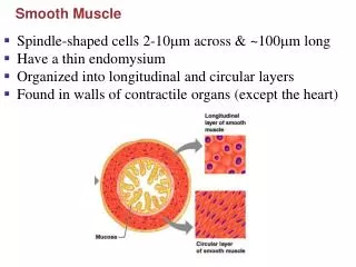
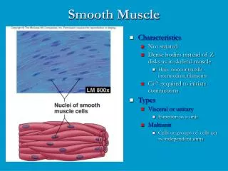
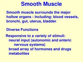
Smooth muscle • Muscle lining vessels & hollow organs, such as the gastrointestinal tract, the bladder, and the uterus, the ureters and the bronchioles. • Sheets of small spindle-shaped (fusiform) cells, connected by special junctions. • Single nucleus (mononucleate). • Contractile units randomly arranged
Special features of Smooth Muscle • Not striated. • Contractile units randomly arranged • NOZline ( Instead Dense Bodies present in the cytoplasm & in the cell membrane ) • Lots of actin, some myosin • T –Tubule absent, hence triads also absent • Caveoli – Analog of T –Tubule • Sarcoplasmic reticulum is poorly developed • Troponin is absent, instead calmodulin is present
“Sidepolar”cross bridges Most of the Myosin filaments have arranged so that the bridges on one side hinge in one direction & those on the other side hinge in the opposite direction that it allows smooth muscle cells to contract as much as 80 per cent of theirlength ● Contractions have longer duration and less tension
The fibrillar contractile apparatus ●Dense bodies serve as an attachment points for the thin filaments ●Intermediate filaments form a cytoskeletal network between them mechanical junctions intermediate filament contractile proteins dense bodies gap junctions
Types of Smooth Muscle Single unit (most): • Clusters of smooth cells, connected by gap junctions. • Activity in one cell spreads to other cells and acts as a single functional unit. e.g. gut, uterus, bladder. Multi-unit:Acts as individual units , no syncytium • e.g. Intrinsic muscles of eye (iris), skin piloerectors, large blood vessels .
Fewer gap junctions assure independent contraction and more precise control.
Single unit 1. Large sheets 2. Low resistance bridges 3. Syncytium Seen in: uterus, urethra, intestine, ureter, urinary bladder 5. Pace maker activity present Multi unit Individual units No bridges Non- syncytial Iris, Ciliary muscles of the eye, piloerector muscle of skin, blood vessels No pace maker activity Single unit & Multi unit SM - Differences
Single unit 6. Rhythmic contraction and relaxations are independent of innervation 7.Stretch causes contraction 8.No tetanic contracton Multi unit Contractions are stimulated only by ANS Stretch cannot cause contraction Irregular tetanic contractions present
IN THE CLINIC Hirschsprung's disease • The enteric nervous system controls many aspects of gastrointestinal function, including motility. • Some children are born without enteric nerves in the distal portion of the colon. • In these children, normal motility of the colon does not occur and severe constipation results. • It can be corrected by surgically removing the portion of the colon that does not contain enteric nerves
Basic steps in contraction of SM Depolarisation of muscle fibres ↓ Release of Ca++ from sarcoplasmic reticulum & from ECF ↓ Ca++ combines with calmodulin ↓ Activation of myosin light chain kinase ↓ Phosphorylation of myosin ↓ Sliding of actin over myosin
Calponin and Caldesmonare other two thin filamentous proteins which in resting state of muscle bind actin and also inhibit myosin ATPasepreventing the interaction of actin and myosin. • Ca2+-calmodulin complex leads to their phosphorylation which in turn releases them from their inhibition.
Q Which statement below most closely describes the role of calcium ions in the control of smooth muscle contraction? (A) Binding of calcium ions to regulatory proteins on thin filaments removes the inhibition of actin-myosin interaction (B) Binding of calcium ions to regulatory proteins associated with thick filaments, specifically calmodulin, activates the enzymatic activity of myosin molecules (C) Calcium ions serve as a direct inhibitor of the interaction of thick and thin filaments (D) A high concentration of calcium ions in the myofilament space is required to maintain muscle in a relaxed state
Relaxation in smooth muscles • Occurs when the intracellular Ca2+ concentration falls. • Mechanisms for fall in intracellular Ca: • Repolarisation of sarcolemmal membrane which closes voltage gated Ca2+ channels • Direct inhibition of Ca2+ channels on sarcolemma by ligands such as cGMP • Inhibition of IP3 production and decreased release of Ca2+ from sarcoplasmic reticulum • SERCA pump • Activation of myosin light chain phosphatase which dephosphorylates myosin light chain leading to inhibition of myosin ATPase.
Relaxation of SM Contraction ↓ Dephosphorylation of myosin ↓ - Relaxation or - sustained contraction due to latch bridgemechanism i.e. Prolonged tonic contraction for hours with little use of energy
Some contractile activity patterns exhibited by smooth muscles
Characteristics of smooth muscle contraction • Slow cycling of myosin cross bridges – less ATPase activity • Less energy required for sustaining muscle contraction • Slowness of onset of contraction and relaxation • Maximum force of contraction is greater • Latch mechanism for prolonged holding of contraction
Electrical properties of SM • RMP (-50mv). It is Unstable leading to spontaneous excitation. • Depolarisationdue to the entry ofCa++mainly and Na+ to a lesser extent. Repolarisation due to delayed K+ efflux & closure ofCa++channels. 3. Sinusoidal waves (Basal electric rhythm) can be recorded from the longitudinal muscles of stomach and intestine. This decides the frequency of peristalsis.
Electrical properties of SM – contd. 4.Action potentials - Spike potentials *appears either on the up going or down going wave of sinusoidal wave * decides the intensity of peristaltic wave - Pacemaker potentials * are generated in multiple foci that shift from place to place * responsible for spontaneous excitation - Plateau type * significance not known
An action potential in smooth muscle can be associated with a slow twitch-like response, and the twitch forces can summate during periods of repetitive action potentials (i.e., similar to tetany in skeletal muscle). Such a pattern of activity is characteristic of single-unit smooth muscle in many viscera
Action potential 0 Ca2+ influx K+ efflux Membrane potential (mV) -45 Threshold of VOC’s -60 200 msec
Relationships between membrane potential (Em) and generation of force (F) in different types of smooth muscle
Mechanical properties of SM 1. Spontaneous activity seen. 2. Maintained state of partial contraction is Tonus or Tone. 3. No correlation between electrical & mechanical events Muscle starts contracting 200msec after the spike – peak contraction is after 500msec
Mechanical properties of SM– contd. 4.Plasticity • Relationship between initial length of muscle fiber and tension • Tension is variable at any given length • Studied in Human urinary bladder • Useful inaccomodating more blood by vessels, more food by stomach & more urine by bladder without a rise in pressure.
Smooth and skeletal muscle mechanicalcharacteristics compared.
Innervation By Autonomic nervous system Sympathetic NS causes Relaxation (Intestine), Contraction (Blood vessels) through Norepinephrine and Epinephrine Parasympathetic NS causes Contraction (Intestine) through Acetyl choline
Other factors that influence SM: • Hormones & drugs: • Adrenaline & Noradrenaline : Similar to SNS • ACh : Similar to PNS • Atropine : Cause relaxation by blocking parasympathetics. • Pilocarpine : Similar to Ach • Estrogen and Progesterone on uterus • Oxytocin at the time of labour • Cold, Stretch & barium ions stimulate contraction. • Hypoxia, hypercapnia, acidosis, K+ cause relaxation