Download
1 / 80
800 likes | 848 Vues
Discover the intricate workings of muscle types, fibers, and neuromuscular junctions, including the mechanisms of muscle contraction and neurotransmitter action. Gain insights into fast-twitch and slow-twitch fibers along with the role of acetylcholine and other neurotransmitters.

E N D
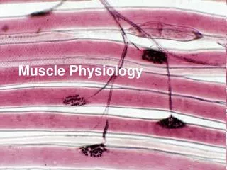
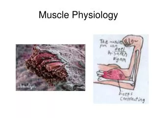
Physiology of Muscle Humaryanto
TIPE OTOT • Otot Skeletal (lurik/striata) • Otot Jantung (lurik/striata) • Otot Polos (polos) (GI, VU, Vascular)
Extrafusal Muscle Fibers • Striate muscle • Force for limb movements • flexion - closes joint • extension - opens joint • Contract or relax ~
OTOT SKELETAL • 40% BB tubuh • Fungsi : mengatur posisi dan gerak rangka • Melekat ke tulang melalui tendo • Origo : perlekatan pada bag. proksimal, bersifat stasioner • Insersio : perlekatan pada bag. distal, bersifat mobil
TIPE OTOT SKELETAL Berdasarkan kecepatan kontraksi dan daya tahan terhadap fatigue. • Fast-twitch glycolitic fibers (putih) • Fast-twitch oxidative fibers (merah) • Slow twitch oxidative fibers (merah) Setiap orang punya 3 tipe otot, tapi berbeda pada komposisi dominan (Jauhari Johan vs John Murray/ Ben Johnson)
Type 1 Fibers • Slow fibers • dark red • slow, sustained contraction • slow to fatigue • Aerobic metabolism • many capillaries & mitochondria • oxygen required for ATP synthesis • myoglobin • gives dark red appearance ~
Type 2b Fibers • Fast fatigable fibers • white fibers • rapid, brief contraction • fast to fatigue • produce about 10x force of Type 1 • Anaerobic metabolism • fewer capillaries & mitochondria • ATP generated by glycolysis • lactic acid buildup ~
Type 2a Fibers • Fast fatigue-resistant fibers • pale red • properties intermediate to types 1 & 2b • rapid, brief contraction • slow to fatigue • produce least force • Aerobic & Anaerobic metabolism • many capillaries & mitochondria ~
Neuromuscular Junction • Synapse between neuron & effector • Cholinergic (ACh) • nicotinic receptors • Motor end-plate • postsynaptic membrane • folds packed with receptors • increased surface area ~
Global view of a neuromuscular junction:1. Axon2. Motor end-plate3. Muscle fiber4. Myofibril
Detailed view of a neuromuscular junction:1. Presynaptic terminal2. Sarcolemma3. Synaptic vesicle4. Nicotinic acetylcholine receptor5. Mitochondrion
Mechanism of action • Upon the arrival of an action potential at the axon terminal, voltage-dependent calcium channels open and Ca2+ ions flow from the extracellular fluid into the motor neuron's cytosol. This influx of Ca2+ triggers excitation-contraction coupling, a biochemical cascade that causes neurotransmitter-containing vesicles to fuse to the motor neuron's cell membrane and release acetylcholine into the synaptic cleft. • Acetylcholine diffuses across the synaptic cleft and binds to the nicotinic acetylcholine receptors that dot the motor end plate. • The receptors are ligand-gated ion channels, and when bound by acetylcholine, they open, allowing sodium and potassium ions to flow in and out of the muscle's cytosol, respectively.
Mechanism of action • Because of the differences in electrochemical gradients across the plasma membrane, more sodium moves in than potassium out, producing a local depolarization of the motor end plate known as an end-plate potential (EPP). • This depolarization spreads across the surface of the muscle fiber into transverse tubules, eliciting the release of calcium from the sarcoplasmic reticulum, thus initiating muscle contraction. • The action of acetylcholine is terminated when the enzymeacetylcholinesterase degrades the neurotransmitter and the unhydrolysed neurotransmitter diffuses away
Neurotransmitters and Neuromodulators • Neuromodulators modify the postsynaptic cell's response to neurotransmitters or change the presynaptic cell's synthesis, release or metabolism of the neurotransmitter. Acetylcholine (Ach) • Major neurotransmitter. Fibers that release ACh are called cholinergic fibers. Acetylcholine is degraded by the enzyme, acetylcholinesterase. Biogenic Amines • Biogenic amines are neurotransmitters containing an amino group. Catecholamines such as dopamine, norepinephrine and epinephrine, serotonin. Nerve fibers that release epinephrine and norepinephrine are called adrenergic and noradrenergic fibers respectively.
Neurotransmitters and Neuromodulators Amino Acid Neurotransmitters • Amino acid neurotransmitters are the most prevalent neurotransmitters in CNS. Glutamate, aspartate GABA (gamma aminobutyric acid), glycine, Neuropeptides • Neuropeptides are composed of two or more amino acids. Neurons releasing neuropeptides are called peptidergic. Beta-endorphin, dynorphin, enkephalins. • Nitric oxide, ATP, adenine also act as neurotransmitters. Neuroeffector Communication • Many neurons of peripheral nervous system end at neuroeffector junctions on muscle and gland cells. Neurotransmitters released by these efferent neurons then activate the target cell.
Muscle Contraction • AP generated in muscle fiber (cell) • Ca++ released from internal stores • Muscle fiber contracts • continues while Ca++ & ATP available • Relaxation • Ca++ sequestered by active transport ~
Muscle Fiber Structure • Multinucleated • fusion of multiple precursor cells • Sarcolemma Excitable membrane • Myofibrils: contractile units • Sarcopasmic reticulum (SR) • sequesters Ca++ • T tubules • AP from sarcolemma to SR • like inside-out axons ~
Miofibril struktur kontraksi otot • 1 Serat otot, tdd: ribuan miofibril • 1 miofibril tdd: Aktin & miosin (protein kontraksi) Troponin & tropomiosin (protein pengatur) Titin & nebulin (protein asessoris besar) • Miosin thick filament, punya kepala Motor protein, E kimia E mekanik, mgd ATP-ase (hidrolisis) • Aktin thin filament, melekat troponin & tropomiosin • Titin molekul elastis (protein terbesar) stabilitas & elastisistas otot • Nebulin penyanggah aktin
Sarcoplasmic Reticulum T tubules Myofibrils Sarcolemma
Myofibril: structure & function • Sarcomeres • repeating sections • Z lines • dividers between sarcomeres • thin filaments anchored to Z lines • actin & troponin • Thick filaments between thin filaments • myosin • Contraction:filaments slide by each other ~
Z line Z line Thin filaments Thick Filaments Sarcomere
KONTRAKSI OTOT • Menghasilkan force / gaya muscle tension • Melawan beban/ load • Memerlukan energi (dari ATP) Pencetus kontraksi otot 1. Neuromuscular junction : Rangsang somatik rangsang listrik 2. Excitation-contraction coupling Potensial aksi signal Ca++ siklus kontr-relaks SIKLUS KONTRAKSI DAN RELAKSASI Sliding filaments theory
Contraction • Excitation-contraction coupling • Myosin “heads” crossbridges w/ actin • Ca++ dependent • binds to troponin, reveals binding site • Myosin head rotates • “ratchets” actin inward ~
Contraction • ATP binds to myosin ---> detachment • cocks myosin ---> binds again • rigor mortis: no ATP fibers remain crosslinked • Repeats as long as Ca++ present • sequestered via active transport ~
SLIDING FILAMENT THEORY • Serat otot memendek (overlapping thick & thin filament) • Sliding aktin terhadap miosin • Gaya dari crossbridge miosin mendorong aktin (power stroke) • Crossbridge miosin mendorong aktin menuju pusat sarkomer • Setelah power stroke kepala miosin melepas aktin untuk mengikat bagian aktin yang lain, demikian seterusnya jadi siklus. Analogi : menarik tambang.
In the absence of calcium ions, tropomyosin blocks access to the mysosin binding site of actin. • When calcium binds to troponin, the positions of troponin and tropomyosin are altered on the the thin flament and myosin then has access to its binding site on actin. • Myosin hydolyzes ATP and undergoes a conformational change into a high-energy state. • The head group of myosin binds to actin forming a cross-bridge between the thick and thin filaments.
Ca+2 Ca++ Ca++ Role of Ca+2 in Muscle Contraction * Actin-binding sites are exposed as a result of Ca+2 binding to troponin complex that causes a conformational shift of tropomyosin
The energy stored by myosin is released, and ADP and inorganic phosphate dissociate from myosin. • The resulting relaxation of the myosin molecule entails rotation of the globular head, which induces longitudinal sliding of the filaments. • When the calcium level decreases, troponin locks tropomyosin in the blocking position and the thin filament slides back to the resting state.
Sliding-Filament Mechanism • Muscle contraction is produced by cross bridge cycles. • A cycle has 4 steps: (1) Energizing of myosin cross bridge A + M•ATP —> A + M*•ADP•Pi (ATP is energizer here) (2) Attachment of cross bridge to a thin filament A + M*•ADP•Pi —> A•M*•ADP•Pi (3) Movement of cross bridge, producing tension A•M*•ADP•Pi —> A•M + ADP + Pi (4) Detachment of cross bridge from thin filament A•M + ATP —> A + M•ATP (ATP is modulator here) • Movement of the cross bridges make the overlapping thick and thin filaments slide past each other (they do not change in length) to produce a contraction.
Actin Myofilament During contraction, calcium binds to troponin Covers actin-binding sites at rest
Cross-bridge Cycle This animation by Mike Geeves, Laboratory of Molecular Biology in the UK and the Cambridge Institute for Medical Research
SIKLUS KONTRAKSI • Rigor state: Kepalamiosinterikat dg molekul G-aktin. • ATP menempelkemiosin, kepalamiosinlepasdariaktin. • Hidrolisis ATP: jadi ADP + Pi (masihmenempel) • Miosinmelekatke G-aktin yang baru, energidaripecahnya ATP, saatadapotensialenergi di kepalamiosinuntuk power stroke. • Pi lepas & power stroke: Kepalamiosinberotasimendorongaktinmendekatipusatsarkomer (crossbridge tilting) • ADP lepas: kepalamiosintetapmelekatkeaktin, siapuntuksiklusberikutbilaada ATP yang baru
Excitation-Contraction Coupling Excitation-Contraction (EC) Coupling: 1. An AP travels down a motor (somatic neuron). 2. The AP causes the release of the neurotransmitter acetylcholine into the synapse at the neuromuscular junction. 3. The acetylcholine binds to the acetylcholine receptors on the muscle fiber and cause an EPSP. 4. If the EPSP reaches threshold, an AP is produced on the sarcolemma of the muscle fiber. Meanwhile, the acetylcholine attached to the receptor is destroyed. 5. The AP travels rapidly along the sarcolemma and enters the fiber at every t-tubule.
Excitation-Contraction Coupling 6. As the AP travels through the t-tubule, it causes the Ca++ gates to open and Ca++ flows from the SR into the sarcoplasm. The Ca++ gates close when the AP ends. 7. The increased [Ca++] in the sarcoplasm results in Ca++ binding to troponin. This induces an allosteric change, the tropomyosin is pulled out of the way and steric inhibition is removed. The result is crossbridges begin to form, rotate and break (provided there is plenty of ATP). 8. Cross-bridge cycling continues as long as sarcoplasmic [Ca++] remains high. 9. However, if the Ca++ gates close, the action of the Ca++ ATPase (pump) begins to predominate and sarcoplasmic [Ca++]] drops. When it drops low enough, the troponin loses its Ca++ and changes shape the next time a crossbridge is not in the way. Steric inhibition is quickly re-established and the muscle contraction is over.
Exitation-Contraction Coupling • Dirangsang oleh asetilkolin/achetylcholine Tahap: • Asetilkolin (Ach) lepas dari motor neuron somatik • Ach merangsang potensial aksi serat otot • PA, m’rsg Ca++ lepas dr Ret.Sarkoplasma • Ca++ me’ikat troponin dan m’rsg kontraksi
DHP: Dihydropiridine Saat PA: Ca 100x Relaksasi: Ca masuk RS krn enzim Ca-ATP-ase
PERIODE KONTRAKSI/ TWITCH • Periode Laten (Antara potensial aksi-kontraksi) 2. Periode kontraksi 3. Periode relaksasi Lama periode kontraksi tergantung tipe otot