Download
1 / 14
140 likes | 370 Vues
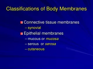
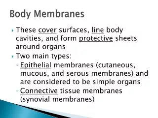
Body Membranes. These cover surfaces, line body cavities, and form protective sheets around organs Two main types: Epithelial membranes (cutaneous, mucous, and serous membranes) and are considered to be simple organs Connective tissue membranes (synovial membranes).

E N D
Body Membranes • These cover surfaces, line body cavities, and form protective sheets around organs • Two main types: • Epithelial membranes (cutaneous, mucous, and serous membranes) and are considered to be simple organs • Connective tissue membranes (synovial membranes)
Cutaneous Membrane • Otherwise known as the skin • Superficial epidermis is composed of a keratinizing (hardening) stratified squamous epithelium • Underlying dermis is mostly dense connective tissue • Since it is exposed to the air, it is considered to be a dry membrane • (More to come later)
Mucous Membranes • Also known as mucosa • Used for absorption and secretion • Epithelium resting on loose connective tissue called lamina propria • Lines all body cavities that open to the exterior, such as the hollow organs of the respiratory, digestive, urinary, and reproductive tracts • They are considered to be wet (moist) membranes
Serous Membranes • Also known as serosa • Simple squamous epithelium resting on a thin layer of areolar connective tissue • Line body cavities that are closed to the exterior
Different areas have different names: • Around the abdominal cavity organs – peritoneum • Around the lungs – pleura • Around the heart - pericardium • Occur in pairs as it folds in on itself • Parietal layer – outer wall (fused to cavity wall) • Visceral layer – inner wall (touches organ) • Space between contains a little bit of a thin, clear liquid called serous fluid (lubricant). • (Remember the balloon and fist model)
Synovial Membranes • Composed of just areolar connective tissue. • Line the fibrous capsules surrounding the joints, small sacs called bursae, and tubeliketendon sheaths • Provide a smooth surface and secrete a lubricating fluid to cushion organs during physical activities
Back to the Cutaneous Membrane And the Integumentary system (the skin and its appendages Some of the basic functions are…
Protects deeper tissue from: • Mechanical Damage • How? It makes a physical barrier. Outer layers has cells that contain keratin, which toughens the cells and has pressure receptors, which alert the body of potential damage • Chemical Damage • How? Once again the keratinized cells block chemicals and pain receptors alert the body
Bacterial Damage • How? Block the bacteria, but also the skin’s secretion are acidic (inhibit growth) and skin cells have phagocytes (eat the bacteria) • Ultraviolet Radiation • How? Contains melanin that offers protection from UV damage • Thermal Damage • How? Contains hot, cold, and pain receptors that alert the body • Desiccation (Drying Out) • How? Contains waterproofing glycolipid and keratin
It also • Aids in heat loss (activates sweat glands and allows blood to flow closer to surface) and heat retention (stops blood from rushing to surface) • Aids in excretion of urea and uric acid (makes up some of your sweat) • Synthesizes vitamin D (uses sunlight to convert some cholesterol molecules to vitamin D)
Structure of the Skin • Epidermis – “top layer” of skin, made up of stratified squamous epithelium that is capable of keratinizing • Dermis – “bottom layer” of skin, made of dense connective tissue • Hypodermis or Subcutaneous tissue – not a layer of skin, but connects skin to organs, mostly adipose tissue and acts as a shock absorber
5 Layers (strata) of the Epidermis • Stratum Basale – lies closest to the dermis and therefore the blood supply, they are constantly dividing and pushing cells up to the surface • Stratum Spinosum – become flatter and fuller of keratin • Stratum Granulosum – keep getting flatter and fuller • Stratum Lucidum – cells die and form this clear layer • Stratum Corneum – cells are completely dead and full of karetin. About the top 20-30 cell layers thick. Layer closest to surface
Regions of the Dermis • Papillary Layer • upper dermal region • Contain fingerlike projections called dermal papillae, which may contain capillary loops to help nourish the epidermis, pain receptors (nerve endings) and touch receptors (Meissner’s corpuscles) • On hands and feet they are arranged in a specific pattern. The result? Fingerprints.
Regions of the Dermis • Reticular Layer • Deepest Layer of the skin • Contains blood vessels, sweat and oil glands, phagocytes, and deep pain receptors called Pacinian corpuscles • Contains both collagen (for toughness and water retention) and elastic fibers (for stretchiness) • Contains blood vessels for heat control • Contains nerves for responsiveness