Photoinduced Cytotoxicity of K562 Cells Using Acridine Orange and Propidium Iodide Staining
100 likes | 248 Vues
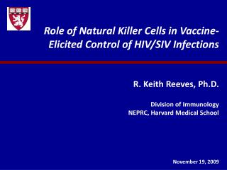
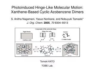
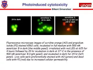
This study examines the photoinduced cytotoxicity effects on K562 cells using fluorescence microscopy. Cells were stained with Acridine Orange (AO) and Propidium Iodide (PI) to differentiate between live (green) and dead (red) cells under various conditions. K562 cells were incubated in full medium with a 500 nM sensitizer for 24 hours in darkness. The middle panel shows cells incubated in the dark, while the right panel displays cells irradiated with red LED light at 625 nm for 4 hours, followed by a 20-hour dark incubation at 37°C.

Photoinduced Cytotoxicity of K562 Cells Using Acridine Orange and Propidium Iodide Staining
E N D
Presentation Transcript
Bilkent Üniversitesi Photoinduced cytotoxicity Fluorescence microscope images of acridine orange (AO) and propidium iodide (PI) stained K562 cells, incubated in full medium with 500 nM sensitizer 3 in dark (the middle panel); irradiated with red LED at 625 for 4 hours followed by 20 hr incubation in dark at 37 oC in the presence of 500 nM sensitizer 3 (right panel); and incubated in dark for 24 hr (left panel). Live cells were preferentially stained with AO (green) and dead cells with PI (red) due to increased cellular permeability.
Bilkent University CNT-derivatization
Bilkent University SWCNTs as carries for PDT reagents
Bilkent University SWCNTs as carriers for PDT reagents • AFM image of compound 1-SWNT. (B) AFM 3D topographic view. • PSIA XE-100E AFM and Multi75AI tip was used in noncontact mode with resonance frequency 75 KHz and force constant 3.0 N/m.
Bilkent University SWCNTs as carriers for PDT reagents
PDT reagents with pH-modulated activity Bilkent University dye DPSB 670 nm dye DPSB + H+
Moore’s Law Bilkent University