Download

1 / 72
760 likes | 1.45k Vues
Carbuncle with Ipsilateral Facial Cellulitis in a Diabetic. Big Group Discussion Group C3B Malig-Marayag. Infections in Diabetics. Diabetic patients have greater frequency and severity of infection due to: Diminished vascularization Hyperglycemia

E N D
Carbuncle with Ipsilateral Facial Cellulitis in a Diabetic Big Group Discussion Group C3B Malig-Marayag
Infections in Diabetics • Diabetic patients have greater frequency and severity of infection due to: • Diminished vascularization • Hyperglycemia • Impairs killing of micro-organisms by neutrophils and macrophages • Interferes with T lymphocyte function • Aids in colonization and growth of a variety of organisms (Candida and other fungal species Oxford Textbook of Medicine, 4th ed. Harrison’s Principles of Internal Medicine, 17th ed.
Cellulitis • Acute suppurative inflammation involving the subcutaneous tissue • Characterized by: • Localized pain, erythema, swelling, heat • Differentiated from erysipelas: • Lesion is not raised • Line between the involved and uninvolved tissue is indistinct Harrison’s Principles of Internal Medicine, 17th ed. Jawetz, Melnick & Adelburg’s Medical Microbiology, 24th ed.
Cellulitis • Mild local erythema and tenderness • Rapidly becomes intense and spreads • Area becomes infiltrated and pits on pressure • Central part may become nodular and develop a vesicle that ruptures and discharges pus and necrotic material • Malaise • Fever and chills Andrews’ Diseases of the Skin: Clinical Dermatology, 10th ed.
Cellulitis • Most commonly caused by indigenous flora • Staphylococcus aureus – usually associated with an abscess, folliculitis, or foreign body • Streptococcus pyogenes – spreads more rapidly; associated with fever and lymphangitis • Bacteria may gain access to the epidermis through: • Cracks in the skin, abrasions, cuts, burns, insect bites, surgical incisions, intravenous catheters Harrison’s Principles of Internal Medicine, 17th ed.
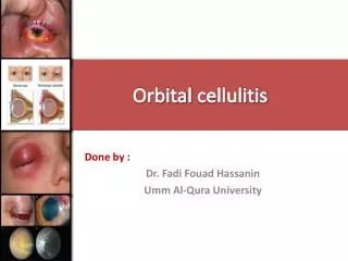
Cellulitis • Associated with predisposing conditions • Streptococcus agalactiae – diabetes mellitus, peripheral vascular disease • Haemophilus influenzae – causes periorbital cellulitis children with sinusitis, otitis media or epiglottitis Harrison’s Principles of Internal Medicine, 17th ed.
Facial Cellulitis • People with certain risk factors are more likely than others to develop facial cellulitis. Facial cellulitis risk factors include: • Problems in the lymphatic system • Upper respiratory infection • Infection of the teeth or middle ear http://skin.emedtv.com/facial-cellulitis/facial-cellulitis-p2.html
Facial Cellulitis • in the facial region the most common portals of entry are dental infections, sinusitis, upper respiratory tract infection, surgery, and trauma Facial cellulitis associated with Pseudomonas aeruginosa complicating ophthalmic herpes zoster Laura Atzori MD1, Caterina Ferreli MD1, Myriam Zucca MD1, Daniela Fanni MD2, and Nicola Aste MD1Dermatology Online Journal 10 (2): 20
Patient: M.E. (55 y/o male), unemployed • Chief complaint: • Painful erythematous swelling on the face
PAST MEDICAL HISTORY • Diabetic for 27 years • Maintained on Gliclazide(Diamicron) for the first 10 years with unrecalled dose taken OD • Insulin maintenance (Humulin N 20 ’u’ in the morning and 15 ‘u’ in the evening) for the past 16 years • Cholecystitis- underwent cholecystectomyin 1996 • Underwent 3 operations for the right eye: • 2003- cataract surgery • 2004- glaucoma-trabeculectomy • 2007- corneal transplant; patient developed graft rejection which led to blindness
FAMILY HISTORY • (+) DM – mother • (+) HPN - father • (+) HPN – brother • (-) Cancer, allergy, stroke
PERSONAL AND SOCIAL HISTORY • Married (with 2 children) • Used to work as a “master cutter” at a tailoring shop until 2003 • Currently unemployed • Occasionally smokes and drinks alcohol • Mixed diet
MEDICATIONS • Insulin (Humulin N 20 ”U” in the morning and 15 “U” in the evening) • Vitamin B complex
REVIEW OF SYSTEMS • No headache, vertigo, syncope • No epistaxis, nasal discharge • No neck stiffness, masses, lymphadenopathy • No tinnitus, ear discharge, loss of hearing • No dyspnea, cough • No chest pain, easy fatigability, nocturnal dyspnea, orthopnea, palpitations
REVIEW OF SYSTEMS • No nausea, vomiting, hematemesis, dysphagia, abdominal pain, diarrhea, constipation, melena, hematochezia • (+) polyphagia, polydypsia, polyuria, (-) dysuria, flank pain, urethral discharge • No joint stiffness, pain, swelling, muscle pain, cramps, weakness, wasting • (+) numbness, tingling sensations on both feet • (+) hyperpigmented scaly plaque on the dorsum of the right foot • No heat-cold intolerance • No pallor, abnormal bleeding, bruising
SEPT 19, 2009 PHYSICAL EXAMINATION ON ADMISSION (SEPT 18, 2009) Conscious, coherent and oriented to time, place and person Vital Signs: BP: 130/70mmHg PR - 109bpm, RR-20cpm, Temp: 36.7°C Weight= 66 kg Height= 170cm BMI=23 HEENT: (+) periorbital swelling, nonhyperemic conjunctivae, anictericsclerae, cornea opaque, OU, retina pigmented OU, lens cannot be assessed, (+) light perception, OD, (+) hand movement, OS No nasoaural discharge, (+) swelling with violaceous discoloration of the lower lip and surrounding skin topped with multiple erosions, crusts and pus, buccal mucosa cannot be assessed NECK: supple neck, no masses, (-) palpable cervical LN,right, LN on the left cannot be assessed due to swelling over the submandibular area, (-) neck vein distension Conscious, coherent and oriented to time, place and person Vital Signs: BP: 120/80mmHg PR - 90bpm, RR-24cpm, Temp: 37.3°C Weight= 66 kg Height= 170cm BMI=23 HEENT: (+) periorbital swelling, nonhyperemic conjunctivae, anictericsclerae, cornea opaque OU No nasoaural discharge (+) swelling with ruptured carbuncle on the left lower lip sorrounded by cellulitis, buccal mucosa cannot be assessed NECK: supple neck, no masses, (-) palpable cervical LN,right, LN on the left cannot be assessed due to swelling over the submandibular area, (-) neck vein distension
PHYSICAL EXAMINATION THORAX & LUNGS: Symmetric chest expansion, (-) retractions (-) lagging, equal vocal tactile fremiti, resonant on all lung fileds, clear breath sounds CARDIAC: Adynamicprecordium, (-) heaves, thrills and lifts, S1>S2 at the apex, S2>S1 at the base, (-) murmurs Pulses full and equal on all extremities ABDOMEN: soft, globular abdomen, non-tender, normoactive bowel sounds; Liver-smooth, non-tender, vertical span- 6 cm at the MCL; spleen & kidneys not palpable EXTREMITIES: Motor strength grade 5/5 on all extremities, no sensory deficits (+) 16X7cm pruritic, hyperpigmented plaque with scaly surface on the dorsum of the right foot and distal third of the leg THORAX & LUNGS: Symmetric chest expansion, (-) retractions (-) lagging, equal vocal tactile fremiti, resonant on all lung fileds, clear breath sounds CARDIAC: Adynamicprecordium, (-) heaves, thrills and lifts, S1>S2 at the apex, S2>S1 at the base, (-) murmurs Pulses full and equal on all extremities ABDOMEN: soft, globular abdomen, non-tender, normoactive bowel sounds; Liver-smooth, non-tender, vertical span- 6 cm at the MCL; spleen & kidneys not palpable EXTREMITIES: Motor strength grade 5/5 on all extremities, no sensory deficits, tingling sensation on both feeth (+)16X7cm pruritic, hyperpigmented plaque with scaly surface on the dorsum of the right foot and distal third of the leg ON ADMISSION (SEPT 18, 2009) SEPT 19, 2009
CLINICAL IMPRESSION • Carbuncle, Left lower lip with Ipsilateral Facial Cellulitis • Diabetes Mellitus, Type 2, Insulin Requiring DIFFERENTIAL DIAGNOSIS: Erysipelas
Cellulitis vs Erysipelas Harrison’s Principles of Internal Medicine 17th ed.
DIAGNOSTIC PLANS: • CBC • CBG • Blood Chemistry • creatinine • Lipid profile • HBA1c • Sodium • Potassium • Arterial Blood Gas
THERAPEUTIC PLANS • ANTIBIOTIC- what? • INSULIN THERAPY ACCORDING TO CBG RESULTS(SLIDING SCALE)/INSULIN DRIP? • DIETARY MANAGEMENT- DIABETIC/RENAL DIET specific diet: kilocalories/day, protein & carbohydrate requirement • HYDRATION-PLAIN NSS (HOW MUCH INFUSION/MIN)
National Diabetes Group and WHO diagnostic criteria for DM: • Fasting plasma glucose level at or above 126 mg/dL (7.0 mmol/L). • Plasma glucose at or above 200 mg/dL (11.1 mmol/L) two hours after a 75 g oral glucose load as in a glucose tolerance test. • Symptoms of hyperglycemia and plasma glucose at or above 200 mg/dL (11.1 mmol/L). Harrison’s 17th Edition
Fasting blood glucose test • The most common test for diagnosis of diabetes. • blood glucose levels are checked after fasting for between 12 and 14 hours. • Patients with fasting glucose levels from 100 to 125 mg/dL (6.1 and 7.0 mmol/L) are considered to have impaired fasting glucose • Patients with diabetes may be asked to delay their diabetes medication or insulin dose until the test is completed. http://ovennewyork.com/diabetes-mellitus-laboratory-tests-or-diagnostic-tests.html
Random blood glucose test • blood glucose levels are checked at various times during the day, and it doesn’t matter when you last ate. • Blood glucose levels tend to stay constant in a person who doesn’t have diabetes. http://ovennewyork.com/diabetes-mellitus-laboratory-tests-or-diagnostic-tests.html
Oral glucose tolerance test (OGTT) • FBS is obtained before the ingestion of a 50- to 200-g glucose load (usual amount is 75 g), • blood samples are drawn at ½, 1, 2, and 3 hours (may be 4- or 5-hour sampling). • Blood samples are checked at regular intervals for two hours. • Glucose tolerance tests are used when the results of the fasting blood glucose are borderline. http://ovennewyork.com/diabetes-mellitus-laboratory-tests-or-diagnostic-tests.html
They are also used to diagnose diabetes in pregnancy (gestational diabetes). • NORMAL: the results of the glucose tolerance test will show that their blood sugar levels fall within the normal range • Patients with plasma glucose at or above 140 mg/dL or 7.8 mmol/L, but not over 200, two hours after a 75 g oral glucose load are considered to have impaired glucose tolerance.
Glycated Hemoglobin (Glycohemoglobin, HbA1c) for Diabetes Mellitus • Measures glycemic control over a 60- to 120-day period by measuring the irreversible reaction of glucose to hemoglobin through freely permeable erythrocytes during their 120-day lifecycle. • While not used for diagnosis, an elevated level of glucose irreversibly bound to hemoglobin of 6.0% or higher (the 2003 revised U.S. standard) is considered abnormal by most labs http://ovennewyork.com/diabetes-mellitus-laboratory-tests-or-diagnostic-tests.html
GOALS OF THERAPY • Eliminate symptoms related to hyperglycemia • Reduce or eliminate the long-term microvascular and macrovascular complications of DM • Allow the patient to achieve as normal a lifestyle as possible Harrison’s 17th Edition
Insulin Insulin vs. hypoglycemics Oral hypoglycemics Should not be used in severly ill patients with type 2 DM Insulin secretagogues: such as sulfonlyureas are effective in individual with relatively recent onset (<5 yrs) of type 2DM Biguanides (metformin): should not be used in patients with renal insufficency [serum creatinine > 133umol/L (1.5mg/dL) in men] Should be discontinued in patients who are severly ill, in patients who can take nothing orally and in thoe receiving radiograohic contrast material α- glucosidase inhibitors (acarbose and miglitol): should not be used in patients with inflammatory bowel disease, gastroparesis or a serum creatinine >177umol/L (2.0mg/dL) Considered initial therapy in type 2 DM, particularly in lean individuals or those with severe weight loss, in individuals with underlying renal or hepatic disease that precludes oral glucose-lowering agents, or in individuals who are hospitalized or acutely ill A long acting insulin (lantus) is best for the patient because if his age etc. since you don’t have to take it often.
Patient education about DM, nutrition and exercise • Patient with DM should receive education about: • Nutrition • Exercise • Care of diabetes during illness • Medications to lower the plasma glucose • Continuing process with regular visits for reinforcement • Diabetes self-management education (DSME) Harrison’s 17th Edition
Diabetes education • Self-monitoring of blood glucose • Urine ketone monitoring (type 1) • Insulin administration • Guidelines for diabetes management during illness • Management of hypoglycemia • Foot and skin care • DM management before, during and after exercise • Risk-factor-modifying activities Harrison’s 17th Edition
Nutrition • Medical Nutrition Therapy (MNT) • Modest caloric reduction • Reduced fat intake • Increased physical activity • Reduction of hyperlipidemia and hypertension Harrison’s 17th Edition
Exercise • CV risk reduction • Reduced BP • Maintenance of muscle mass • Reduction in body fat and weight loss • Lowering plasma glucose • Increasing insulin sensitivity *ADA recommends 150 min/week (distributed over at least 3 days) Harrison’s 17th Edition
Monitoring the level of glyceminc control • Plasma glucose measurements by the patient and assessment of long-term control by the physician • Measurement of A1C and review of the patient’s self-measurement of plasma glucose Harrison’s 17th Edition
Self-monitoring of Blood Glucose • Standard of care in diabetes management and allows the patient to monitor his/her blood glucose at any time • Glucose monitors can rapidly and accurately measure glucose in small amounts of blood (3-10 µL obtained from the fingertip *individuals with type 2 DM who are taking insulin should utilize SMBG more frequently than than those on oral agents Harrison’s 17th Edition
Assessment of Long-term Glycemic Control • Measurement of glycated hemoglobin • Plasma glucose is consistently elevated = increase in nonenzymatic glycation of hemoglobin • Reflects the glycemic history over the previous 2-3 months Harrison’s 17th Edition
DIAGNOSTIC PLANS FOR CELLULITIS CT scan with contrast of the paranasal and neck area pharyngolaryngoscopy Blood culture Aspiration CT scan
*As clinically indicated; • †Ulcerated lesions should be cleaned and debrided before having wound base swabbed; • ‡Most useful if vesicle/bullae or fluid abscess present; • §Seek out bone trauma and air fluid levels; • ¶Indications –neurological deficits, vision nonassessable, proptosis/deteriorating acuity or colour/bilateral edema/ophthalmoplegia, no improvement after 24 h and swinging pyrexia not resolving within 36 h (for head only); • **Only if central nervous system involvement suspected
Diagnosis • Based on appearance of the skin and patient history • Drainage from an abscess or weeping wound associated with cellulitis should be sent for culture and sensitivities. • Material from needle aspiration of inflamed skin or skin biopsy can be cultured in cases of cellulitis without purulence, abscess, or a necrotic • Indications for blood cultures include significant fever and chills, severe immunocompromise, periorbital cellulitis, and cellulitis superimposed on lymphedema. • A polymorphonuclear leukocytosis is often present with cellulitis; a complete blood cell count and differential may help gauge the severity of infection and the hematologic response.