Spinal Nerves
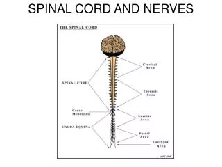
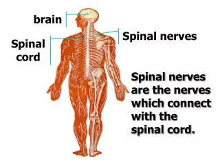
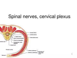
Spinal Nerves. Lecture 4. Location and Structure. There are 31 pairs of spinal nerves, each pair numbered according to the level of the spinal cord from which it arises. The cervical nerves are labeled C1 – C8 The thoracic nerves are labeled T1 – T12 The lumbar nerves are labeled L1 – L5

Spinal Nerves
E N D
Presentation Transcript
Spinal Nerves Lecture 4
Location and Structure • There are 31 pairs of spinal nerves, each pair numbered according to the level of the spinal cord from which it arises. • The cervical nerves are labeled C1 – C8 • The thoracic nerves are labeled T1 – T12 • The lumbar nerves are labeled L1 – L5 • The sacral nerves are labeled S1 – S5 • There is only one coccygeal nerve
Each nerve is attached to the spinal cord by two roots: the dorsal root and the ventral root. • On each dorsal root is a marked swelling of gray matter called the dorsal root ganglion, which contains the cell bodies of the sensory neurons. • A ganglion is any collection of nerve cell bodies located outside of the CNS. Fibers from sensory receptors throughout the body lead to these dorsal root ganglia. • The ventral roots of the spinal nerves are a combination of motor fibers that supply muscles and glands. The cell bodies of these neurons are located in the ventral horns of the spinal cord. • Because the dorsal and ventral roots are combined to form the spinal nerve, all spinal nerves are mixed nerves.
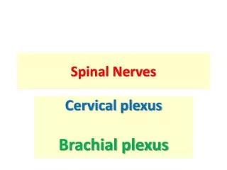
Branches of the Spinal Nerves • Each spinal nerve continues only a short distance away from the spinal cord and then branches into small posterior divisions and large anterior divisions. • The larger anterior branches interlace to form networks called plexuses, which then distribute branches to the body parts. • The three main plexuses are • The cervical plexus (nerves C1 – C5) – supplies motor impulses of the neck and receives sensory impulses from the neck and back of the head. The phrenic nerve, which activates the diaphragm, arises from this plexus.
The brachial plexus (nerves C6 – T1) – sends numerous branches to the shoulder, arm, forearm, wrist,and hand. The radial nerve emerges from the brachial plexus. • The lumbosacral plexus (nerves T12 – S5) – supplies nerves to the pelvis and legs. The larges branch in this plexus is the sciatic nerve, which leaves the dorsal part of the pelvis, passes beneath the gluteus maximus, and extends down the back of the thigh. At its beginning, it is nearly 1-inch thick, but is soon branches to the thigh muscles; near the knee, it forms two subdivisions that supply the leg and the foot.
Dermatomes • Sensory neurons from all over the skin, except for the face and scalp, feed information into the spinal cord through the spinal nerves. • The skin surface can be mapped into regions that are supplied by a single spinal nerve. • These regions are called dermatomes. • Stimulation of the skin within a given dermatome is carried over the corresponding spinal nerve. • For example, the spinal nerve that carries stimulation for the dermatome of the shoulder region is C-4, the knee is L-3, and the toes are S-1.