Download
1 / 20
230 likes | 964 Vues
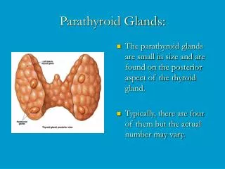
Morphology pf parathyroid adenoma - lies in close proximity to the thyroid gland or in an ectopic site (the mediastinum ) b. Invested by a capsule is almost invariably confined to single gland

E N D
Morphology pf parathyroid adenoma - lies in close proximity to the thyroid gland or in an ectopic site (the mediastinum) b. Invested by a capsule • is almost invariably confined to single gland d. and the remaining glands are somewhat shrunken, as a result of feedback inhibition by elevated serum calcium d. . Most parathyroid adenomas weigh between 0.5 and 5 g.
On microscopic examination, parathyroid adenomas a- Are composed predominantly of chief cells b- A few nests of larger oxyphil cells also are also present. c- A rim of compressed, non-neoplastic tissue, separated by a fibrous capsule, is visible at the edge of the adenoma. d- Cells with pleomorphic nuclei may be seen (endocrine atypia) and must not be taken as a sign of malignancy. e. Mitotic figures are rare f. with inconspicuous adipose tissue
2. Parathyroid hyperplasia • Is a multiglandular process - In some cases, however, enlargement may be grossly apparent in only one or two glands, complicating the distinction between hyperplasia and adenoma. - The combined weight of all glands rarely exceeds 1.0 g .
3. Parathyroid carcinomas : a. Affects one gland b.- Consist of irregular nodules that sometimes exceed 10 g in weight c. A a dense, fibrous capsule encloses the mass. Note - The diagnosis of carcinoma based on cytologic detail is unreliable, and invasion of tissues and metastasis are the only definitive criteria - Local recurrence occurs in one third of cases, - More distant dissemination occurs in another third
I. Skeletal changes in primary hyperparathyroidism a. Osteitisfibrosacysticacharacterized by 1. Increased osteoclastic activity, resulting in erosion of bone and mobilization of calcium salts, mainly in the metaphyses of long tubular bones. 2. Bone resorption is accompanied by increased osteoblastic activity and the formation of new bone 3. In more severe cases the cortex is grossly thinned .4. The marrow contains increased amounts of fibroustissue accompanied by hemorrhage and cysts
b. Brown tumors of hyperparathyroidism • Aggregates of osteoclasts,,and hemorrhage occasionally form masses that may be mistaken for neoplasms II. Kidney changes a. PTH-induced hypercalcemia favors the formation of urinary tract stones (nephrolithiasis) s b. Calcification of the renal interstitium (nephrocalcinosis) III.Metastatic calcification may be seen in the stomach, lungs, myocardium, and blood vessels.
b. Secondary Hyperparathyroidism - Is caused by any condition causing a chronic decrease in the serum calcium level, because low serum calcium leads to compensatory overactivity of the parathyroids. - Renal failure is the most common cause 1. Chronic renal insufficiency causes decreased phosphate excretion, which in turn results in hyperphosphatemia. and the elevated serumphosphate levels depress serum calcium levels and so stimulate parathyroid gland activity
2.Loss of renal substances reduces the availability of α1-hydroxylase enzyme necessary for the synthesis of the active form of vitamin D, which in turn reduces intestinal absorption of calcium Gross- • The parathyroid glands are hyperplastic and called secondary hyperplasia. Clinically a- Are dominated by those related to chronic renal failure
b.- Bone abnormalities (renal osteodystrophy) are less severe than those seen in primary type c.- Serum calcium remains near normal because compensatory increase in PTH levels sustains serum calcium. d- The metastatic calcification of blood vessels (secondary to hyperphosphatemia) occasionally may result in significant ischemic damage to skin and other organs-a process sometimes referred to as calciphylaxis
c.Tertiary hyperparathyroidism - In a minority of patients, parathyroid activity may become autonomous and excessive, with resultant hypercalcemia-a process termedtertiary hyperparathyroidism - Parathyroidectomy may be necessary to control the hyperparathyroidism in such patients
HYPOPARATHYROIDISM: The major causes are:. • Surgically induced hypoparathyroidism: - Inadvertent removal of parathyroids during thyroidectomy. b. Congenital absence: • This occurs in conjunction with thymicaplasia (Di George syndrome) and cardiac defects c. Autoimmune hypoparathyroidism : - This is a hereditary polyglandular deficiency
syndrome arising from autoantibodies to multiple endocrine organs(parathyroid, thyroid, adrenals, and pancreas). Clinical manifestations - Are secondary to hypocalcemia and include: a. Increased neuromuscular irritability (tingling, muscle spasms, facial grimacing, and sustained carpopedal spasm or tetany), b. Cardiac arrhythmias, and, on occasion, increased c. Seizures.
I. AdrerenocorticalHyperfunction (Hyperadrenalism) 1. Hypercortisolism (Cushing Syndrome) - In clinical practice, most cases are caused by the administration of exogenous glucocorticoids (Iatrogenic) - The remaining cases are endogenous and caused by one of the following A. Primary hypothalamic-pituitary diseases associated with hypersecretion of ACTH (Cushing disease) - Accounts for 70% of cases of spontaneous, endogenous Cushing syndrome .
Occurs most frequently during young adulthood (the 20s and 30s) and mainly affecting women Causes of Cushing disease a. ACTH-producing microadenoma (most common) b. Corticotroph cell hyperplasia which may be: a. Primary b. secondary to excessive ACTH release by a hypothalamic (CRH)-producing tumor
- The adrenal glands in Cushing disease show bilateral nodular cortical hyperplasia secondary to the elevated levels of ACTH ("ACTH-dependent" Cushing syndrome). - The cortical hyperplasia, in turn, is responsible for the hypercortisolism
B. Primary adrenal hyperplasia and neoplasms - Are responsible for about 10% to 20% of cases of endogenous Cushing syndrome and this form is called ACTH-independent Cushing syndrome, or adrenal Cushing syndrome and its biochemical hallmark is elevated levels of cortisol with low serum levels of ACTH - In most cases, adrenal Cushing syndrome is caused by a unilateral adrenocortical neoplasm, which may be either benign (adenoma) or malignant (carcinoma).
Note- • The overwhelming majority of adrenal hyperplasia are ACTH-dependent, and primary cortical hyperplasia of the adrenal cortices is a rare cause of Cushing syndrome C. Secretion of ectopic ACTH by nonpituitary tumors - Accounts for about 10% of cases of Cushing syndrome mostly caused by small cell carcinoma of the lung, - The adrenal glands undergo bilateral hyperplasia due to elevated ACTH, but the rapid downhill course of patients with these cancers cuts short the adrenal enlargement
MORPHOLOGY of the pituitary in Cushing syndrome Crooke hyaline change : - Results from high levels of glucocorticoids, and in this condition, the normal basophilic cytoplasm of the ACTH-producing cells is replaced by homogeneous slightly basophilic material - This alteration is the result of the accumulation of intermediate keratin filaments in the cytoplasm.
Changes in adrenal in cases of Cushing syndrome: • Cortical atrophy : - If the syndrome results from exogenous glucocorticoids ,suppression of endogenous ACTH results in bilateral cortical atrophy, due to a lack of stimulation of the zonafasciculata and reticularis by ACTH, - The zonaglomerulosa is of normal thickness because it functions independently of ACTH