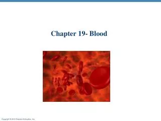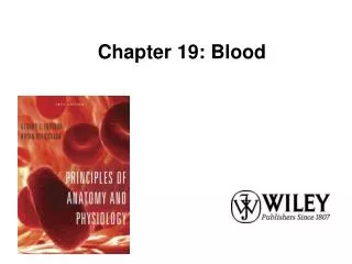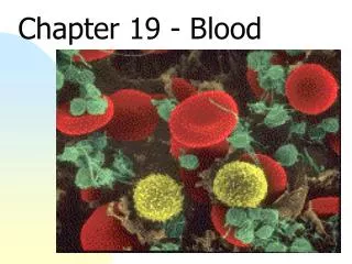
Lecture 19, Blood
Lecture 19, Blood. Lecturer: Dr. Ebadi Room P313 Phone: (718) 260-5285 E-Mail: ibarjis@citytech.cuny.edu. Learning Objectives. List the components of the cardiovascular system and explain the major functions of this system. Describe the important components and major functions of the blood

Lecture 19, Blood
E N D
Presentation Transcript
Lecture 19, Blood Lecturer: Dr. Ebadi Room P313 Phone: (718) 260-5285 E-Mail: ibarjis@citytech.cuny.edu
Learning Objectives • List the components of the cardiovascular system and explain the major functions of this system. • Describe the important components and major functions of the blood • List the characteristics and functions of red blood cells. • Describe the structure of hemoglobin and indicate its functions. • Discuss red blood cell production and maturation.
Learning Objectives • Explain the importance of blood typing and the basis for ABO and Rh incompatibilities. • Categorize the various white blood cells on the basis of structure and function. • Describe the structure, function and production of platelets. • Describe the reaction sequences responsible for blood clotting.
The cardiovascular system • Provides a mechanism for rapid transport of nutrients, waste products, respiratory gases and cells
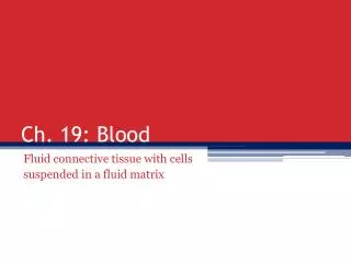
Functions and Composition ofBlood • Fluid connective tissue • Functions include • Transporting dissolved gases, nutrients, hormones, and metabolic wastes • Regulating pH and ion composition of interstitial fluids • Restricting fluid loss at injury sites • Defending the body against toxins and pathogens • Regulating body temperature by absorbing and redistributing heat
The composition of blood • Plasma and formed elements comprise whole blood. • Plasma elements include blood cells: • Red blood cells (RBC) • White blood cells (WBC) • Platelets • Can fractionate whole blood for analytical or clinical purposes
The Composition of Whole Blood Fresh whole blood for testing in a lab is usually collected from a superficial vein. When checking the efficiency of gas exchange, it may be necessary to draw a blood sample from an artery
The Composition of Whole Blood The chief difference between plasma and interstitial fluid involves the concentration of dissolved oxygen and proteins.
Hemopoiesis • Process of blood cell formation • Hemocytoblasts are circulating stem cells that divide to form all types of blood cells • Whole blood from anywhere in the body has roughly the same temperature (38ºC), pH (7.4) and viscosity. • Bright red color if taken from artery • Dull red color if taken from vein
Plasma • Accounts for 46-63% of blood volume • 92% of plasma is water • Higher concentration of dissolved oxygen and dissolved proteins than interstitial fluid
Plasma proteins • more than 90% are synthesized in the liver • Albumins are the most abundant plasma proteins • 60% of plasma proteins • Responsible for viscosity and osmotic pressure of blood
Additional Plasma Proteins • Globulins • ~35% of plasma proteins • Include immunoglobins which attack foreign proteins and pathogens • Include transport globulins which bind ions, hormones and other compounds • Fibrinogen • Converted to fibrin during clotting • Are necessary for blood clotting • Removal of fibrinogen leaves serum
Red Blood Cells Abundance of RBCs • Erythrocytes (RBC) account for slightly less than half the blood volume, and 99.9% of the formed elements. • Hematocrit measures the percentage of whole blood occupied by formed elements • Commonly referred to as the volume of packed red cells
Structure of RBCs • Biconcave disc, providing a large surface to volume ration • Shape allows RBCs to stack, bend and flex • RBCs lack organelles • Typically degenerate in about 120 days.
Hemoglobin • Molecules of hemoglobin account for 95% of the proteins in RBCs • Hemoglobin is a globular protein, formed from two pairs of polypeptide subunits • Each subunit contains a molecule of heme which reversibly binds an oxygen molecule • Damaged or dead RBCs are recycled by phagocytes
RBC life span and circulation • Replaced at a rate of approximately 3 million new blood cells entering the circulation per second. • Replaced before they hemolyze • Components of hemoglobin individually recycled • Heme stripped of iron and converted to biliverdin, then bilirubin • Iron is recycled by being stored in phagocytes, or transported throughout the blood stream bound to transferrin
RBC Production • Erythropoeisis = the formation of new red blood cells • Occurs in red bone marrow • Process speeds up with in the presence of EPO (Erythropoeisis stimulating hormone) • RBCs pass through reticulocyte and erythroblast stages
Blood types • Determined by the presence or absence of surface antigens (agglutinogens) • Antigens A, B and Rh (D) • Antibodies in the plasma (agglutinins) • Cross-reactions occur when antigens meet antibodies
The White Blood Cells Leukocytes • Have nuclei and other organelles • Defend the body against pathogens • Remove toxins, wastes, and abnormal or damaged cells • Are capable of amoeboid movement (margination) and positive chemotaxis • Some are capable of phagocytosis
Types of WBC Granular and agranular • Granular leukocytes • Neutrophils – 50 to 70 % total WBC population • Eosinophils – phagocytes attracted to foreign compounds that have reacted with antibodies • Basophils – migrate to damaged tissue and release histamine and heparin
Types of WBC • Agranular leukocytes • Agranular leukocytes are formed inred bone marrow. • Agranular leukocytes include: • Monocytes - become macrophage • Lymphocytes – includes T cells, B cells, and NK cells
Differential count • Indicates a number of disorders • Leukemia = inordinate number of leukocytes
WBC Production • Granulocytes and monocytes are produced by bone marrow stem cells • Divide to create progenitor cells • Stem cells may originate in bone marrow and migrate to peripheral tissues • Several colony stimulating factors are involved in regulation and control of production
The Origins and Differentiation of Formed Elements Animation: The origins and differentiation of blood cells (see tutorial) Figure 19.12
Platelets • Flattened discs • Circulate for 9-12 days before being removed by phagocytes
Platelet functions • Transporting chemicals important to clotting • Forming temporary patch in walls of damaged blood vessels • Contracting after a clot has formed
Platelet production (thrombocytopoiesis) • Megakaryocytes release platelets into circulating blood • Rate of platelet formation is stimulated by thrombopoietin, thrombocyte-stimulating factor, interleukin-6, and Multi-CSF
Hemostasis • Prevents the loss of blood through vessel walls • Three phases – • Vascular phase • Platelet phase • Coagulation phase
Hemostasis • Vascular phase • Local blood vessel constriction (vascular spasm) • Platelet phase • Platelets are activated, aggregate at the site, adhere to the damaged surfaces
Coagulation phase • Factors released by platelets and endothelial cells interact with clotting factors to form a clot • Extrinsic pathway • Intrinsic pathway • Common pathway • Suspended fibrinogen is converted to large insoluble fibrin fibers
Clot retraction • Final phase of healing • Platelets contract and pull the edges of the vessel together
Fibrinolysis • Clot gradually dissolves through action of plasmin • Activated form of plasminogen • Clotting can be prevented through the use of drugs that depress the clotting response or dissolve existing clots • Anticoagulants include heparin, coumadin, aspirin, dicumarol, t- PA, streptokinase, and urokinase
You should now be familiar with: • The components of the cardiovascular system and its major functions. • The important components and major functions of the blood. • The characteristics and functions of red blood cells. • The structure of hemoglobin and its functions. • Red blood cell production and maturation.
You should now be familiar with: • The importance of blood typing and the basis for ABO and Rh incompatibilities. • The various white blood cells. • The structure, function and production of platelets. • The reaction sequences responsible for blood clotting.