Neonatal Liver Biopsy
430 likes | 932 Vues
Neonatal Liver Biopsy. Dr Claire Bowen Consultant Paediatric Pathologist. Topics Covered. Neonatal jaundice Indications for biopsy Handling of biopsy in the laboratory Histological assessment of the liver biopsy Patterns of liver disease with examples Biliary atresia A1AT

Neonatal Liver Biopsy
E N D
Presentation Transcript
Neonatal Liver Biopsy Dr Claire Bowen Consultant Paediatric Pathologist
Topics Covered • Neonatal jaundice • Indications for biopsy • Handling of biopsy in the laboratory • Histological assessment of the liver biopsy • Patterns of liver disease with examples • Biliary atresia • A1AT • Cystic fibrosis • Neonatal hepatitis • Metabolic
Neonatal Jaundice • Common • 60% term and 80% of preterm babies develop jaundice in first week of life • 10% breast fed babies still jaundice at 1 month • Usually harmless • High concentrations of conjugated hyperbilirubinaemia can cause permanent brain damage (kernicterus) • Prolonged jaundice can be a sign of underling serious liver disease (conjugated bilirubinaemia >25 umol/L) • Early recognition and prompt treatment essential • Phototherapy • Kasai portoenterostomy • Metabolic screening • Transplant
Indications Biopsy indicated • Conjugated hyperbilirubinaemia • Jaundice persisting beyond 2 weeks (3 weeks in preterm babies) • Dark urine • Pale stools • Total parenteral nutrition in the context of intestinal failure • Assessment of rejection post-transplant • Tumour Biopsy not indicated • Unconjugated hyperbilirubinaemia • Physiological • Sepsis • Haemolysis • Liver failure • Coagulopathic • Limited contribution to aetiology • Usually see explanted liver
Handling Procedure risk - general anaesthetic, bleeding Need maximum amount of information from biopsy Light microscopy Snap frozen tissue for metabolic cases Electron microscopy for storage disorders Dry tissue for copper
H&E • Number and size of tissue cores • Portal tracts • Number • Presence/absence of bile ducts • Bile duct proliferation • Inflammation • Fibrosis • Parenchyma • Giant cell transformation • Rosetting of hepatocytes • Haematopoiesis • Storage cells • Central veins • Vascular flow abnormalites • Inflammation in rejection
Special stains • Connective tissue stains to assess fibrosis • EVG – mature fibrosis/pericellular fibrosis • Reticulin – cell plates, acute collapse • Trichrome – tends to overestimate fibrosis • Orcein - Copper associated protein and Hep B • Perls to assess iron • PAS/DPAS – glycogen, storage cells and Alpha-1-Antitrypsin globules
1) Biliary Features • Fibrosis • Fibrous portal tract expansion • Bridging fibrosis • Lobular pattern of cirrhosis • Ductular proliferation • Ductular bile plugging • Periportal copper-associated protein • Variable giant cell change • Haematopoiesis
Differential diagnosis Extrahepatic biliary atresia Alpha-1-antitrypsin (mimic) Total parenteral nutrition Cystic fibrosis – eosinophilic secretions in bile duct and fatty change
Biliary Atresia Rare - 1 in 17000 in UK Presents in first few weeks 50 cases a year with normal antenatal scans 20% other anomalies (cardiac, polysplenia) Lumen of biliary tree obliterated with obstruction to bile flow Progressive liver damage – cirrhosis 5 categories of postulated aetiology –> Inflammatory, Developmental, Vascular, Environmental and Viral
Alpha-1 Antitrypsin Defective A1AT protein Defective production of A1AT leading to decreased A1AT activity in the blood and lungs Deposition of excessive abnormal A1AT protein in liver cells. Mimics – Can see biliary pattern or giant cell pattern PAS positive globules within hepatocytes – not identified in first 3 months Immuno for A1AT
Cystic Fibrosis • Liver disease 5% in CF patients • Fibrosis • Cholestatsis • Fatty change • Biliary features • Mucin in bile ducts characteristic but not always seen
2) Neonatal / giant cell hepatitis Largely normal portal tracts Hepatocyte disarray and collapse Florid giant cell change Rosetting of hepatocytes Cholestasis Extramedullary haematopoiesis May see storage cells Not usually fibrotic
Differential diagnosis Idiopathic with spontaneous recovery Infection Metabolic Storage A1AT
3) Paucity of bile ducts • Bile duct proper lacking • Need at least 10 portal tracts (1 in 10 miss bile duct normally) • Abberent periportal cytokeratin 7 expression • Normally stains biliary epithelium • Stains periportal hepatocytes where there is duct loss
Differential diagnosis • Syndromic • Alagilles syndrome • Non-syndromic • CMV infection • A1AT • Cystic fibrosis • Chronic rejection
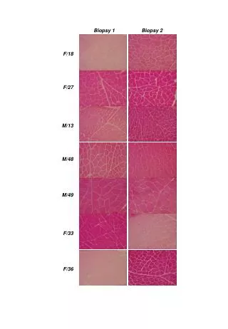
Alagille syndrome Characteristic facial features – triangular face Heart problems Bile ducts seen in early biopsies Progressive bile duct loss/absence Fibrosis Abberant Cytokeratin 7 staining in periportal hepatocytes
Early features Late Features
4) Bland cholestasis Canalicular cholestasis No ductular reaction or bile plugging Minimal parenchymal changes +/- Fibrosis
Pitfalls in children • Copper-associated protein present in babies up to 3 months (periportal) • Small amounts of periportal iron present at birth • Fat not generally seen, metabolic conditions should be considered • Hepatocyte plates 2 cells thick until 5/6 years • Erythropoiesis stops approx. 36 weeks gestation
Neonatal Haemochromatosis Severe form of iron overload Starts to accumulate in utero - can cause fetal death Liver failure Massive necrosis – collapse Iron +++ Usually diagnosed on lip biopsy – iron storage in salivary glands