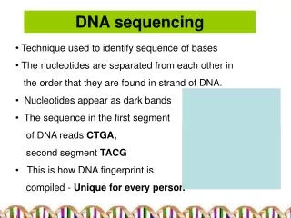
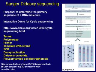
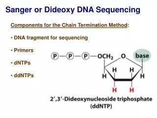
DNA sequencing by the Sanger method
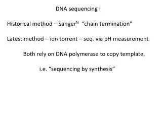
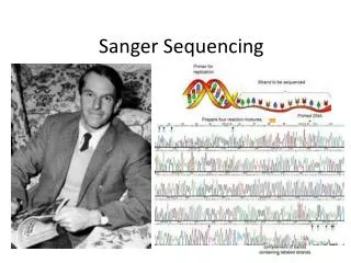
DNA sequencing by the Sanger method. The standard DNA sequencing technique is the Sanger method, named for its developer, Frederick Sanger, who shared the 1980 Nobel Prize in Chemistry. This method begins with the use of special enzymes to synthesize fragments of DNA that terminate

DNA sequencing by the Sanger method
E N D
Presentation Transcript
DNA sequencing by the Sanger method The standard DNA sequencing technique is the Sanger method, named for its developer, Frederick Sanger, who shared the 1980 Nobel Prize in Chemistry. This method begins with the use of special enzymes to synthesize fragments of DNA that terminate when a selected base appears in the stretch of DNA being sequenced. These fragments are then sorted according to size by placing them in a slab of polymeric gel and applying an electric field -- a technique called electrophoresis. Because of DNA's negative charge, the fragments move across the gel toward the positive electrode. The shorter the fragment, the faster it moves. Typically, each of the terminating bases within the collection of fragments is tagged with a radioactive probe for identification.
DNA sequencing example Problem Statement: Consider the following DNA sequence (from firefly luciferase). Draw the sequencing gel pattern that forms as a result of sequencing the following template DNA with ddNTP as the capper. atgaccatgattacg... Solution: Given DNA template: 5'-atgaccatgattacg...-3' DNA synthesized: 3'-tactggtactaatgc...-5'
DNA sequencing example Given DNA template: 5'-atgaccatgattacg...-3' DNA synthesized: 3'-tactggtactaatgc...-5' Gel pattern: +-------------------------+ lane ddATP | W | | || | lane ddTTP | W | | | | | | lane ddCTP | W | | | | lane ddGTP | W || | | +-------------------------+ Electric Field + Decreasing size where "W" indicates the well position, and "|" denotes the DNA bands on the sequencing gel.
A sequencing gel This picture is a radiograph. The dark color of the lines is proportional to the radioactivity from 32P labeled adenonsine in the transcribed DNA sample.
Reading a sequencing gel You begin at the right, which are the smallest DNA fragments. The sequence that you read will be in the 5'-3' direction. This sequence will be exactly the same as the RNA that would be generated to encode a protein. The difference is that the T bases in DNA will be replaced by U residues. As an example, in the problem given, the smallest DNA fragment on the sequencing gel is in the C lane, so the first base is a C. The next largest band is in the G lane, so the DNA fragment of length 2 ends in G. Therefore the sequence of the first two bases is CG. The sequence of the first 30 or so bases of the DNA are: CGTAATCATGGTCATATGAAGCTGGGCCGGGCCGTGC.... When this is made as RNA, its sequence would be: CGUAAUCATGGUCAUAUGAAGCUGGGCCGGGCCGUGC.... Note that the information content is the same, only the T's have been replaced by U's!.
Translating the DNA sequence The order of amino acids in any protein is specificed by the order of nucleotide bases in the DNA. Each amino acid is coded by the particular sequence of three bases. To convert a DNA sequence First, find the starting codon. The starting codon is always the codon for the amino acid methionine. This codon is AUG in the RNA (or ATG in the DNA): GCGCGGGUCCGGGCAUGAAGCUGGGCCGGGCCGUGC.... Met In this particular example the next codon is AAG. The first base (5'end) is A, so that selects the 3rd major row of the table. The second base (middle base) is A, so that selects the 3rd column of the table. The last base of the codon is G, selecting the last line in the block of four.
Translating the DNA sequence This entry AAG in the table is Lysine (Lys). Therefore the second amino acid is Lysine. The first few residues, and their DNA sequence, are as follows (color coded to indicate the correct location in the codon table): Met Lys Leu Gly Arg … ... AUG AAG CUG GGC CGG GCC GUG C.. This procedure is exactly what cells do when they synthesize proteins based on the mRNA sequence. The process of translation in cells occurs in a large complex called the ribosome.
Automated procedure for DNA sequencing A computer read-out of the gel generates a “false color” image where each color corresponds to a base. Then the intensities are translated into peaks that represent the sequence.
High-throughput seqeuncing:Capillary electrophoresis The human genome project has spurred an effort to develop faster, higher throughput, and less expensive technologies for DNA sequencing. Capillary electrophoresis (CE) separation has many advantages over slab gel separations. CE separations are faster and are capable of producing greater resolution. CE instruments can use tens and even hundreds of capillaries simultaneously. The figure show a simple CE setup where the fluorescently-labeled DNA is detected as it exits the capillary. Sheath flow Laser Focusing lens Sheath flow cuvette Beam block Collection Lensc Collection Lensc PMT filter
Sieving matrix for CE It is not easy to analyze DNA in capillaries filled only with buffer. That is because DNA fragments of different lengths have the same charge to mass ratio. To separate DNA fragments of different sizes the capillary needs to be filled with sieving matrix, such as linear polyacrylamide (acrylamide polymerized without bis-acrylamide).This material is not rigid like a cross- linked gel but looks much like glycerol. With a little bit of effort it can be pumped in and out of the capillaries. To simulate the separation characteristics of an agarose gel one can use hydroxyethylcellulose. It is not much more viscous then water and can easily be pumped into the capilliaries.