Download
1 / 19
200 likes | 417 Vues
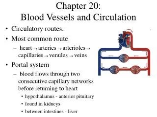
Nervous system. Chapter 20 Meninges and blood vessels of the brain and spinal cord, and the cerebrospinal fluid. The menings of brain and spinal cord The blood vessels of brain and spinal cord The cerebrospinal fluid and its circulation Blood brain barrieer.

E N D
Nervous system Chapter 20 Meninges and blood vessels of the brain and spinal cord, and the cerebrospinal fluid • The menings of brain and spinal cord • The blood vessels of brain and spinal • cord • The cerebrospinal fluid and its • circulation • Blood brain barrieer
The meninges of brain and spinal cord the meninges of spinal cord spinal dura mater epidural space subdural space spinal arachnoid mater subarachnoid space terminal cistern spinal pia mater denticulate ligament
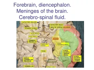
cerebullar meninges From outside to inside: cerebral dura mater cerebral arachnoid mater cerebral pia mater
cerebral dura mater cerebral falx tentorium of cerebellum cerebellar falx diaphragma sellae
Dural sinuses superior sagittal sinus inferior sagittal sinus straight sinus transverse sinus sigmoid sinus
cavernous sinus superior petrosal sinus inferior petrosal sinus ⑴ visual tract hypophysis ⑶ cavernous sinus ⑷ ⑸ • The structures which go through the interior wall of cavernous sinus: • internal carotid artery ⑴ • abducent nerve ⑵ ⑵ ⑴ ⑹ • The structures which go through the lateral wall of cavernous sinus: • from upper to down • oculomotor nerve ⑶ • trochlear nerve ⑷ • trigeminal ophthalmic nerve branch(V1) ⑸ • superior maxillary nerve branch(V2)⑹
the afflux of blood in sinuses of dura mater: superior sagittal sinus inferior sagittal straight confluens transverse sigmoid internal sinus sinus sinus sinus sinus jugular vein cavernous superiorpetrosal sinus sinus inferior petrosal sinus
cerebral arachnoid mater subarachnoid space subarachnoidal cistern interpeduncular cistern cerebellomedullary cistern superior cistern of quadrigeminal bodies chiasmatic cistern cisterna pontis arachnoid granulations cerebral pia mater Thecerebral pia mater and its blood vessels as well as ependymal epithelium of this region constitute chorioid tela commonly. The choroid plexus is the main structure of producing cerebrospinal fluid.
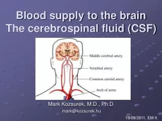
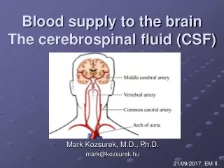
The blood vessels of brain and spinal cord cerebullar blood vessels cerebullar artery internal carotid artery anterior cerebral artery and anterior communicating artery middle cerebral artery anterior choroidal artery posterior communicating artery
vertebral artery basilar artery The principal branches of vertebral artery are: anterior spinal artery, posterior spinal artery posterior inferior cerebellar artery the principal branches of basilar artery anterior inferior cerebellar artery auditory artery(Internal auditory artery) pontine arteries superior cerebellar artery posterior cerebral artery arterial circle of Willis(Willis circle)
cerebullarvein Don’t go with artery, and infuse to sinuses of dura mater via many pathways, finally return via internal jugular vein. Superficial group superior cerebral vein (above lateral sulcus) inferior cerebral vein (below lateral sulcus) Middle cerebral vein superficial middle cerebral veins deep middle cerebral vein
Deep group internal cerebral veins great cerebral vein(Galen vein)
myelonic blood vessel myelonicartery posterior spinal artery anterior spinal artery vasocorona
myelonic vein is more and thicker than artery. The anterior vein and posterior vein are confluenced by the veinlet of spinal cord,and influxed internal vertebral vein plexus of epidural space by vena radix anterior and posterior radical vein.
cerebrospinal fluid and its circulatin Thecerebrospinal fluid is produced by choroid plexus of every cerebral ventricle, which is colorless and transparent fluid and filled with ventricular system、central canal of spinal cord and subarachnoid space. The cerebrospinal fluid play a role of buffering shock, protection, transporting metabolic product and regulating intracranial pressure.
The route ofcerebrospinal fluid circulation choroid plexus of lateral ventricle produce cerebrospinal fluid interventricular foramen third ventricle of cerebrum(choroid plexus produce cerebrospinal fluid) aqueduct of midbrain fourth ventricle of cerebrum(choroid plexus produce cerebrospinal fluid) lateral aperture、median aperture subarachnoid space arachnoid granulations superior sagittal sinus
blood-brain barrier blood-brain barrier structural basement: capillary endothelium basal lamina of blood capillary gelatinous membrane blood-CSF barrier structural basement: Zona occludens exist on the interspaceal top of choroid plexus epithelial cell.
thestructural basement of cerebrospinal fluid-blood-brain barrier the structural basement: ependymal epithelium、cerebral pia mate and gelatinous membrane below meninx vasculosa Theblood-brain barrier can protect the brain and spinal cord from the influence of physical and chemical factor in or out of environment, thus to maintain relative steady state.