Osmoregulation
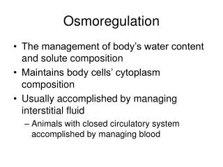
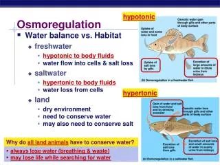
Osmoregulation. Controlling the Internal Environment. terms. Hypertonic: exo-smosis Hypotonic: endosmosis These have led to structural adaptations and physiological adaptations in order to maintain the balance of water and solutes.


Osmoregulation
E N D
Presentation Transcript
Osmoregulation Controlling the Internal Environment
terms • Hypertonic: exo-smosis • Hypotonic: endosmosis • These have led to structural adaptations and physiological adaptations in order to maintain the balance of water and solutes. • Excretion : expulsion of waste products of metabolism. Ex urea, uric acid ammonia
Functions of excretory system • Toxins and other substances that are ingested and absorbed but are not fully metabolized Ex: betain, certain drugs • Excess water, produced by cell respiration and absorbed from food in the gut • Excess salt absorbed from food in the gur • Nitrogenous wasted Ex urea
Functions of Excretory System • Regulate body fluids • Volume, pH, composition • Remove metabolic wastes from blood • Control rate of RBC formation • Regulate blood pressure • Regulate absorption of calcium
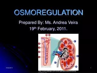
The structure of Kidney Papilla Pyramid Cortex Medulla Hilum Column Calyx Pelvis Ureter
Structure of kidney • The kidney has a bean-shaped structure, each kidney has concave and convex surfaces. The concave surface, the renal hilum, is the point at which the renal artery enters the organ, and the renal vein and ureter leave. The kidney is surrounded by tough fibrous tissue, the renal capsule
The kidney is approximately 11–14 cm in length, 6 cm wide and 4 cm thick. • The substance, or parenchyma of the kidney is divided into two major structures • : superficial is the renal cortex and deep is the renal medulla • Grossly, these structures take the shape of 8 to 18 cone-shaped renal lobes, each containing renal cortex surrounding a portion of medulla called a renal pyramid (of Malpighi)
Between the renal pyramids are projections of cortex called renal columns (of Bertin).Nephrons, the urine-producing functional structures of the kidney, span the cortex and medulla. • The initial filtering portion of a nephron is the renal corpuscle located in the cortex, which is followed by a renal tubule that passes from the cortex deep into the medullary pyramids. • Part of the renal cortex, a medullary ray is a collection of renal tubules that drain into a single collecting duct • The tip, or papilla, of each pyramid empties urine into a minor calyx, minor calyces empty into major calyces and major calyces empty into the renal pelvis which becomes the ureter.
Blood supply • The kidneys receive blood from the renal arteries, left and right, which branch directly from the abdominal aorta. Despite their relatively small size, the kidneys receive approximately 20% of the cardiac output
Kidney : structure • Innervation • The kidney and nervous system communicate via the renal plexus, whose fibers course along the renal arteries to reach the kidney. Input from the sympathetic nervous systemtriggers vasoconstriction in the kidney, thereby reducing renal blood flow
Functions • Acid-base homeostasis Acid-base homeostasis • Two organ systems, the kidneys and lungs, maintain acid-base homeostasis, which is the maintenance of pH around a relatively stable value. The kidneys contribute to acid-base homeostasis by regulating bicarbonate(HCO3-) concentration.
Osmolality regulation • Any significant rise in plasma osmolalityis detected by the hypothalamus which communicates directly with the posterior pituitary gland. An increase in osmolality causes the gland to secrete antidiuretic hormone (ADH), resulting in water reabsorption by the kidney and an increase in urine concentration. The two factors work together to return the plasma osmolality to its normal levels.
ADH binds to principal cells in the collecting duct that translocateaquaporins to the membrane allowing water to leave the normally impermeable membrane and be reabsorbed into the body by the vasa recta, thus increasing the plasma volume of the body
Ultra structure of Nephron PCT Bowman’s capsule DCT Vasa recti Glomerulus Ascending tubule Descending Tubule Collecting duct
Structure of Nephron • Bowmans capsule- Bowman’s capsule: Blood is filtered under high pressure (ultrafiltration). Wastes plus some useful molecules plus some water are filtered into the nephron • Glomerulus • Filtration of blood • Water • Electrolytes • Glucose • Urea.
Function of Renal Corpuscle • Proximal convoluted tubule: useful molecules plus most of water are selectively reabsorbed into the blood • Loop of Henle: alters salt concentration in the medulla to aid re-absorption of water from the collecting ducts
Formation of urine • Collecting duct: kidney can reabsorb water from here and return it to the blood according to the body’s demands under the influence of anti diuretic hormone • Urine containing wastes dissolved in a small volume of water
urea • The urea forms the major constituent of urine. It is formed from the de-amination of amino acids. The urea formation takes place in the liver.
Formation of urine • Urine is produced in the kidneys from water and wastes extracted from the blood • Blood is supplied to the kidneys by the renal arteries which branch off the aorta. The kidneys and are drained by the the renal veins into the inferior vena cava
Function of Tubules • Reabsorption of glucose • Proximal convoluted tubule • Reabsorption of water • PCT, descending tubule, collecting duct • Reabsorption of salts • PCT, ascending tubule, distal con. tub. • Reabsorption of urea • PCT, collecting duct
Selective absorption • In selective reabsorption glucose, amino acids and glucose are reabsorbed from the filtrate back into the blood • In the collecting duct kidney can reabsorb water and return to the blood according to the body’s needs.
Ultrafiltration (UF) is a variety of membrane filtration in which hydrostatic pressure forces a liquid against a semi permeable membrane. Suspended solidsand solutes of high molecular weight are retained, while water and low molecular weight solutes pass through the membrane.
In biological terms, ultra filtration occurs at the barrier between the blood and the filtrate in the renal corpuscle or Bowman's capsule in the kidneys. The Bowman's capsule contains a dense capillary network called the glomerulus. Blood flows into these capillaries through a wide afferent arteriole and leaves through a narrower efferent arteriole. The blood pressure inside these capillaries is high because
The renal artery contains blood at very high pressure which enters the glomerulus via the short afferent arteriole. • The efferent arteriole has a smaller diameter than the afferent arteriole. • The high pressure forces small molecules such as water, glucose, amino acids sodium chloride and urea through the filter, from the blood in the glomerular capsule across the basement membrane of the Bowman's capsule and into the nephron. This type of high pressure filtration is ultra filtration. The fluid formed in this way is called glomerular filtrate
Glomerular pressure is about 75 millimeters of mercury (10 kPa). It is opposed by osmotic pressure(30 mmHg, 4.0 kPa) and hydrostatic pressure (20 mmHg, 2.7 kPa) of solutes present in capsular space. This difference in pressure is called effective pressure(25 mmHg)(3.3 kPa).
Glomerular filtration rate • Glomerular filtration rate (GFR) is the volume of fluid filtered from the renal glomerular capillaries into the Bowman's capsule per unit time.. • Glomerular filtration rate (GFR) can be calculated by measuring any chemical that has a steady level in the blood, and is freely filtered but neither reabsorbed nor secreted by the kidneys. • The rate therefore measured is the quantity of the substance in the urine that originated from a calculable volume of blood. The GFR is typically recorded in units of volume per time, e.g. milliliters per minute ml/mi. Compare to filtration fraction • There are several different techniques used to calculate or estimate the glomerular filtration rate (GFR or eGFR).
Osmoregulation • The water content of the body can vary depending on various factors. Hot weather and physical activity such as exercise make us sweat and so lose body fluids. • Drinking tends to be at irregular intervals when socially convenient. This means that sometimes the body has too little water and needs to conserve it and sometimes too much water and needs to get rid of it. Most of the control of water conservation takes place in the distal and collecting tubules of the nephrons under control of anti-diuretic hormone, (ADH), sometimes called vasopressin. • This hormone is released by the posterior pituitary under control of the hypothalamus in the mid-brain area. The hypothalamus monitors the water content of the blood. If the blood contains too little water (indicating dehydration) then more ADH is released. If the blood contains too much water (indicating over-hydration) then less ADH is released into the blood stream
dehydration • Athletes who want to loose weight can take drugs to interfere with ADH. Less water is reabsorbed, more is lost in the urine and the body looses weight • HANGOVER- Alcohol reduces the effect of kidney. Water is reabsorbed and the body becomes dehydrated. Dehydrated brain cells cause a headache.
dialysis • A dialysis machine is artificial kidney. It takes the patient’s blood and ‘ cleans’ it. Wastes diffuse out of the blood across a partially permeable membrane into a fluid that is constantly renewed. In this way urea is removed from the blood
A dialysis machine • Blood is taken from artery leaves body under pressure. • Blood returns to the body through vein (since blood in vein is at low pressure) • Chamber removes blood clots and re-warms blood to body temperature so body is not shocked when blood returns. • Dialysis fluid has a compostion which means that urea and salts diffuse into it from the blood but useful solutes and water do not. • Diffusion of urea out of blood is aided by countercurrent flow of plasma and dialysis fluid- they flow in opposite directions maintaining the concentration gradient.