Download
1 / 14
230 likes | 942 Vues
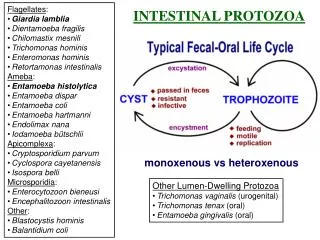
Balantidium coli, Giardia lamblia , and trichomonas . Continue in protozoa. Balantidium coli:. It’s uncommon parasite of humans. It commonly infects pigs and has a worldwide distribution. Two stages can be seen:

E N D
Balantidium coli, Giardialamblia, and trichomonas. Continue in protozoa
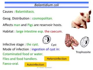
Balantidium coli: • It’s uncommon parasite of humans. • It commonly infects pigs and has a worldwide distribution. • Two stages can be seen: • The trophozoite: large, easily seen oval shaped ciliate with a rapid revolving motility. • The cyst is : large, round, thick walled, it’s the infective stage. Cilia is common but not clearly seen in cyst.
Macronucleus stains well with iodine and other stains. • A large macronucleus may be seen “kidney shape”. A very small micronucleus lies close to the macronucleus but not clearly appear. • Habitat: Large intestine. • Disease: Balantidiasis with dysentery-abdominal pain, mucoid bloody diarrhea with tenesmus. • Complications: Intestinal: haemorrhage, appendicitis, peritonitis. • Extra intestinal: amoeba can migrate and infect other organs (liver, lung, skin, brain). • Diagnosis: stool examination.
Balantidium coli: Cyst Trophozoite
Cyst Trophozoite
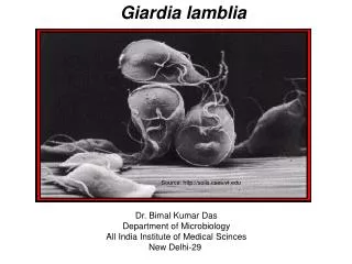
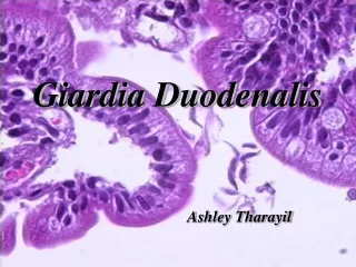
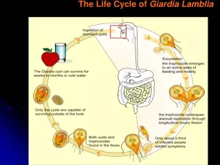
GiardiaLamblia: • In tropical and subtropical areas. • Seen in children more than adults. • Have 2 stages: cyst and trophozoite. • Cyst: oval in shape, with central Axostyle, and 2 nuclei. In direct method may seen without nuclei at 40X. • Trophozoite: pear shape, “monkey face” fragile stage not seen clearly during processing.
Can cause autoinfection. • Infective sate is the cyst. • Habitat: small intestine. • Disease: Giardiasis: abdominal pain, diarrhea, gases and anorexia for 1-2 weeks. • Complications “chronic stage”: malabsorption of fat (steatorrhea) produce pale coloration of the stool. • Diagnosis: stool examination.
Giardialamblia: Cyst Trophozoite
Giardialamblia: Cyst Trophozoite
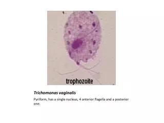
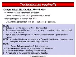
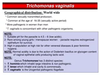
Trichomonas: • Either trichomonasvaginalis or tichomonashominis. • No cystic form in trichomonas, there is trophozoite stage, and so it considered as the infective stage. • Trophozoite is ovoid or pear-shaped. • Has a short undulating membrane. • Has 4 anterior flagella and a fifth running along the outer margin of the undulating membrane. • Has a big vesicular nucleus.
Habitat: in female vagina and cervix, in male urethra. • Diagnosis: in female: vaginal swab or secretions or urine. In male: prostatic secretions or urine. • To differentiate between trichomonasvaginalis and hominis this is by the specimen.