Pathogenic Microorganism Detection Test
Pathogenic Microorganism Detection Test . Object. Understand the principle and the basic procedure of bacterial culture and identification 2. Understand the principle 、 method and result report of antimicrobial susceptibility testing (Kirby-Bauer method) . Diagnostic bacteriology procedures .

Pathogenic Microorganism Detection Test
E N D
Presentation Transcript
Object • Understand the principle and the basic procedure of bacterial culture and identification 2. Understand the principle、method and result report of antimicrobial susceptibility testing (Kirby-Bauer method)
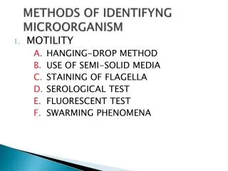
Diagnostic bacteriology procedures • 1、Direct microscopy: • 1)、wet mounts methods • 2)、stain: • A、Gram stain • B、Acid-fast stain : Mycobacterium • tuberculosis • C、Indian ink stain : Cryptococcus • neoformans
2. Culture • Aerobic bacteria • Facultatively anaerobic bacteria culture • Obligately anaerobic bacteria • Isolated colonies ---- “pure culture”
3. Identification • 1) Biochemical identification • 2) Serologic identification • 3) Molecular techniques
4. Antimicrobial susceptibility testing (AST) • Kirby-Bauer disc agar diffusion method (K-B method) : Material Principle Procedure(watch some pictures) Result
The Principle of Kirby-Bauer method • the bacterium is swabbed across an agar plate • paper discs containing antibiotics are placed • a gradient of antibiotic surrounding the disk is formed • incubation • zones of inhibition of bacterial growth may be present • measure the diameter of the zone • determine the sensitivity of the organism to the drug.
Materials 1. Strain: Escherichia coli 2. Medium: Mueller-Hinton agar (d=90mm, h=4mm) 3. Paper discs containing different antibiotics :Ceftazidime (CAZ) 、Ceftriaxone (CRO) 、Aztreonam (AZT) 、Cefotaxime (CTX) 、Gentamicin (GM) 、Tetracycline (TE)
Result significance • S : susceptible, to indicate that the bacteria can be inhibited by the normal dose of antibiotics. • I : intermediate , to indicate that the bacteria can be inhibited by the high dose of antibiotics. • R : resistant , to indicate that the bacteria can not be inhibited by the antibiotics.
Notice 1. Strict sterile operation to prevent contamination. 2. The distance between two paper discs must larger than 24 mm, at the most 6 paper discs can be placed onto a 90~100 mm flat plate. 3. Exactly measure the diameter of zones of inhibition.
Discussion • When MRSA or MRSCON has been detected, which antibiotics can be chose in clinical therapy? • MRS: methicillin-resistant staphylococci • MRSA: methicillin-resistant staphylococcus aureus MRSCON: methicillin-resistant coagulase- negative staphylococcus