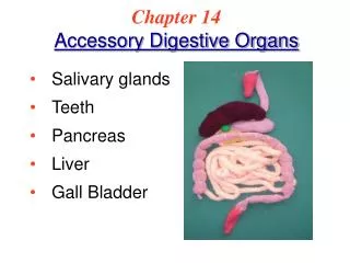
Digestive Glands: Salivary Glands, Pancreas, Liver
510 likes | 535 Vues
Explore the structures and functions of salivary glands, pancreas, and liver in the digestive system. Learn about exocrine and endocrine pancreas, hepatic lobule, and more.

Digestive Glands: Salivary Glands, Pancreas, Liver
E N D
Presentation Transcript
---small gland: fundis gland, small intestinal gland ---large gland: salivary gland, pancreas, liver
1) General structure of exocrine gland ---Capsule: CT, which separate the parenchyma into lobules ---lobule:
① acinus: a. serous acinus: structure: • pyramidal or cuboidal • round basally-located N • apical: zymogen granule-acidophilic • basal: basophilic-RER, ribosome function: secret salivary amylase(淀粉酶)
b. mucous acinus: structure: • pale-stained, slight-blue, flattened N against the basal membrane • mucinogen granule • Golgi, RER, mitochondria
c. mixed acinus: several serous cell attached to mucous acinus – serous demilune
②duct: • intercalated duct: • simple squamous epi. • simple cuboidal epi. • connect with acinus
b. striated duct (secretory duct): intralobule • simple columnar epi. : • tall cell, acidophilic • round N, located at apical part • longitudinal striation: plasma membrane infolding
c. interlobular duct and major duct : simple columnar epi. or pseudostratified ciliated columnar epi.
2) structural feature of each salivary gland a. parotid gland: -serous -long intercalated duct, short striated duct
b. submandibular gland: -mixed( mainly serous, less mucous and mixed) -short intercalated duct, long striated duct
c. sublingual gland: -mixed(mainly mucous, more serous and mixed) -more demilunes, no intercalated duct, under-developed striated duct
胰腺-图 2. Pancreas
---capsule: CT, septa ---parenchyma: /exocrine pancreas /endocrine pancreas
1)exocrine pancreas ---acinus: serous
centroacinar cell: small, pale cell with round or ovoid N, derived from epithelial cell of intercalated duct
---duct: long intercalated duct ---function: secret pancreatic liquid 1-2L/D, PH 7.8-8.4 Digestive enzymes: trypsinogen(胰蛋白酶原), amylase(淀粉酶) ,lipase(脂酶) and chymotrypsinogen(糜蛋白酶原),
2) endocrine pancreas( pancreas islet) ---170.000-200.000, constitute about 1% of total pancreas volume ---75-500 um ---HE: cells arranged into cord with CT rich in fenestrated cap.
a. A cell: • 20%. Large polygonal in shaped, peripheral-distributed • EM: secretory G: large, 190-310 nm, round with dense core • function: secret glucogon(高血糖素) - 29 amino acid residues protein ↓ glycogen→ glucose
b. B cell: • 75%, small, centrally-distributed • EM: secretory G: different diameter, 225-375 nm with one or several dense core • function: secret insulin - 51 amino-acid residues
c. D cell: • 5%, ovoid, fusiform, peripheral-distributed, between A, B cells • EM: -gap junction with A,B cell -secretory G: large, 190-370nm, low-density core • function: secrete somatostatin(生长抑素) to inhibit the secreting of A, B, PP cell
d. PP cell: • EM: secrete granule: small, 110-170 nm • function: secrete pancreatic polypeptide (胰多肽)to inhibit the secreting of pancreatic liquid, movement of viscera and contraction of gall bladder
e. D1 cell: 2-5%, peripheral, irregular EM: small, 140-190 nm, function: secrete VIP (vasoactive intestinal peptide,血管活性肠肽) f. C cell: undifferentiated cell
肝-图 3. Liver
---largest, 2% of body weight ---capsule: DCT, insert into parenchyma to separate the parenchyma into hepatic lobule ---hepatic lobule ---portal area
1) hepatic lobule: basic structural unit ---500,000-1,000,000 ---2 mm long, 1 mm in across D ---polygonal (irregular) ---structure: • central vein • hepatic plate: radiating arranged • hepatic sinusoid
① Central vein: • small vein: endothelium + CT • 45 um • receive the blood from sinusoids
② Hepatic plates ---formed by single layer of hepatocytes
a. hepatocyte: LM: polygonal, 20-30 um eosinophilic N: -large, pale, round, centrally- located -1/4 binucleate
EM: • mitochondria: 1000-2000, 20% total volume • RER: involve in the synthesis of albumin, fibrinogen, clotting factor, lipoprotein and complement protein • SER: contain enzymes- oxidoreductase氧化还原酶 (oxidase, reductase), hydrolase, transterase, synthetase, involve in the formation of bile and the metabolism of adipose, glucose and hormones
Golgi apparatus: involve in -formation of bile -process, condense and storage of proteins -formation of lysosome • Lysosome: involve in phagocytosis activity and metabolism of bilirubin(胆红素) • microbody: -round, 0.2-1.0 um -contain catalase (过氧化氢酶)and peroxidase(过氧化物酶) • inclusions: glycogen, lipid droplet, pigment
b. bile canaliculus: ---cell membrane of adjacent hepatocytes depress to form a tubular system between hepatocytes
---structure: • silver preparation: dark-brown colored network • 0.5-1um • Microvilli • tight junction, desmosome
③ Hepatic sinusoid ---space between hepatic plates ---structure:
9-12 um endothelial cell: fenestrated, gap, plasmalemmal vesicles -liver macrophage (Kupffer cell) -large granular lymphocyte: NK cell
Perisinusoidal space: Disse space - narrow space between endothelial cell and hepatocytes • 0.4 um width; blood plasma • microvilli; RF • fat-storing cell:
fat-storing cell: -irregular, with processes -EM: large lipid droplets, RER, mito, Golgi -function: storage of vitamin A(E,K), synthesis of collagen
The three kinds of different surfaces of hepatocytes ---face adjacent cell each other: 55% ---face the sinusoids: 35% ---form bile canaliculus: 10%
2) portal area ---areas(triangle-shaped or irregular-shaped) where adjacent hepatic lobules meet ---contains CT and several ducts
a. interlobular arteries: • branches of hepatic A • small A: endothelium + 3-4 layers of SM b. interlobular vein: • branches of portal vein • small vein: endothelium + less CT and single SM c. interlobular bile duct: • simple cuboidal or low columnar epi.
3) Blood circulation of liver hepatic A →interlobular A →terminal hepatic arteriole portal V→interlobular V→terminal portal venule → hepatic sinusoid →central vein→sublobular V →hepatic V→inferior vena cava ) {
Blood circulation of liver 门V 小叶间V 终末门微V 血窦 小叶下V 中央V 肝A 小叶间A 终末肝微A 肝V
4) Passage of bile • 肝细胞 胆小管 闰管 小叶间胆管 左右肝管 肝总管 胆囊管 胆囊 或经胆总管到十二指肠