Download
1 / 40
400 likes | 435 Vues
Learn about the types of radiation emitted by nuclear bombs and their effects on the environment and human health. Understand dosimetry units and measurements, radiation detection, and related terminology.

E N D
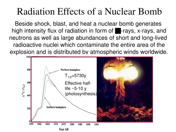
Radiation Effects of a Nuclear Bomb Beside shock, blast, and heat a nuclear bomb generates high intensity flux of radiation in form of -rays, x-rays, and neutrons as well as large abundances of short and long-lived radioactive nuclei which contaminate the entire area of the explosion and is distributed by atmospheric winds worldwide. T1/2=5730y Effective half- life ~5-10 y (photosynthesis)
14C distribution + nuclear test related 14C production
Nuclear Bomb related Radiation Production The units rad (rem) are a measure of radiation exposure!
Classical Unit: 1 Curie [Ci] Modern Unit: 1 Becquerel [Bq] Monitoring radiation intensity The so-called dosimetry units (rad, rem) determine the amount of damage radioactive radiation can do to the human body. They depend on the kind and nature of the incident radiation (X-rays, -rays, -particles, -particle, or neutrons). It also depends on the energy loss of the particular radiation and the associated ionisation effects in the human body material.
Amount of energy E deposited by radiation into body part of mass m. unit Rad or Gray Dose: Radiation independent dose Q is normalization factor which accesses the individual body damage done by the particular kind of radiation Unit Rem or Sievert = × H Q D Equivalent Dose: Radiation Exposure & Dosimetry Photons: Q=1 Neutrons: E<10keV Q=5 Neutrons: E>10keV Q=15 Protons: Q=5 Alphas : Q=20
UNITS OF RADIATION MEASUREMENT Dosage units: The Sievert (Gray) is a measure of biological effect. 1 Gray (Gy) = 1 Joule/kg (Energy/mass) 1 Sievert (Sv) = Gray x Q, where Q is a "quality factor" based on the type of particle. Q for electrons, positrons, and x-rays = 1 Q = 3 to 10 for neutrons, protons dependent upon the energy transferred by these heavier particles. Q = 20 for alpha particles and fission fragments. Converting older units: 1 rad = 1 centigray = 10 milligrays ( 1 rad = 1cGy = 10 mGy ) 1 rem = 1 centisievert = 10 millisieverts ( 1 rem = 1cSv = 10 mSv ) Nominal background radiation absorbed dose of 100 mrad/year = 1 mGy/yr. Nominal background radiation dose biological equivalent of 100 mrem/year = 1mSv/yr. Occupational whole body limit is 5 rem/yr = 50 mSv/yr. 2.5 mrem/hr or 25 uSv/hr is maximum average working level in industry. Exposure rate from Naturally Occurring Radioactive Material; an empirically derived conversion factor for Ra-226 decay series: 1.82 microR/ hour = 1 picoCurie/gram.
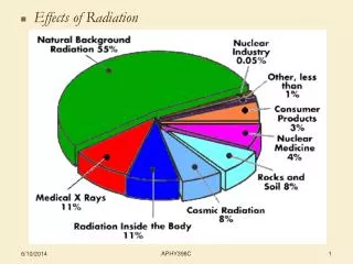
Exposure to Natural and Man-made Radioactivity Total average annual dose: H ≈ 250-300 mrem Tobacco contains -emitter 210Po with T1/2=138.4 days. Through absorption in the bronchial system smoking adds 280 mrem/year to the annual dose of US population Average annual dose from nuclear bomb test fallout Hfo≈ 0.06 mrem.
Cosmic Ray Bombardment Low energy CR • Cosmic Rays origin from: • solar flares; • distant supernovae; Spectrum of CR High energy CR
Cosmic Rays in High Altitude Earth is relatively protected from cosmic rays through atmosphere shield; typical exposure is H=3.2 mrem/h. Mountain climbers and airline crews and passengers are exposed to higher doses of radiation. Dose doubles every 1500 m in height. At 10 km height dose is about 100 times sea-level dose H=0.32mrem/h. • Example: Total dose H: • after 10h of flight: • H=3.2 mrem, • for round trip: • H=6.4 mrem • Frequent flyer with about • 10 transatlantic flights/year • H=64 mrem/year. • Compare to natural dose (~200 mrem/y) !
Au g-activity ObservableEffects! Wife’s ring with ground level dose! 8 times more dose Husband’s ring with transatlantic high altitude dose
b+ b- 40K g 40Ar 40Ca Long lived 40K Radioactivity 40K has a half-life of T1/2=1.28·109 years its natural abundance is 0.021 % Potassium decay to Argon 40K 40Ar
Internal Glowing On average, 0.27% of the mass of the human body is potassium K of which 0.021% is radioactive 40K with a half-life of T1/2=1.25·109 [y]. Each decay releases an average of Eavg= 0.5 MeV - and -radiation, which is mostly absorbed by the body but a small fraction escapes the body. Calculate, how many radioactive 40K atoms are in your body system!
(8.54 Example: 40K Calculate the absorbed body dose over an average human lifetime of t = 70 y for this source of internal exposure.
Prompt Release of Radiation • Nuclear bomb causes sudden release of a high flux on: • -rays E=h≈1-10 MeV electromagnetic waves • x-rays E=h≈1-100 keV electromagnetic waves • -radiation 4He nuclei • -radiation electrons and positrons • neutrons neutrons • heavy radioactive species (cause for delayed radiation) The prompt radiation is absorbed in the surrounding Atmosphere according to exponential absorption law I0is the initial intensity and is the attenuation coefficient determined by the interaction probability of radiation with molecules and atoms in air.
Alpha Beta Gamma Neutron Absorption probability Attenuation coefficient depends on energy and nature of particle, medium and interaction probability. High Coulomb scattering probability for charged particles, causes high absorption probability, results in short range! Main component gammas & neutrons 1 m Concrete
Spread of prompt & secondary -radiation Neutrons originated secondary radiation by inelastic neutron scattering as well as by neutron capture on nitrogen isotopes in the surrounding air. Secondary -production enhances radiation flux and radiation extension.
128Te 129Xe 130Te 127I 128Sn 129Sn 130Sn 125Te 126Te 127In 128In 129In 126Cd 127Cd 128Cd 126Ag 127Ag Fissionproducts Production of neutron-rich radioactive isotopes in the mass 80-130 range which decay by - decay or by - delayed neutron emission Back to stable isotopes. Decay time scale depends On the associated half-lives which determine the flux and time scale for delayed radiation exposure. e.g. 126Ag(-,n)125Cd vs 126Ag(-)126Cd
100 10 1 activity (in %) 0.1 0.01 0.001 0.001 0.01 0.1 1.0 10.0 100.0 1000.0 days Decline by the “rule of seven” This rule states that for every seven-fold increase in time following a fission detonation (starting at or after 1 hour), the radiation intensity decreases by a factor of 10. Thus after 7 hours, the residual fission radioactivity declines 90%, to one-tenth its level of 1 hour. After 7·7 hours (49 hours, approx. 2 days), the level drops again by 90%. After 7·2 days (2 weeks) it drops a further 90%; and so on for 14 weeks. The rule is accurate to 25% for the first two weeks, and is accurate to a factor of two for the first six months. After 6 months, the rate of decline becomes much more rapid.
Studies of impact of ionizing radiation on the human body - Hiroshima - US-Japanese teams medical tests, autopsies, human organ analysis, on-site radioactivity measurements … autopsy
Hiroshima radiation spread data Primary ray originated low dose of <100 rad near the hypocenter, secondary -ray originated dose of >100 rad within 1500 m radius
Irradiation External Contamination Internal Contamination * * Radiation Exposure Types * * * * * *
Schematic Model of Radionuclide Uptake Intake: Inhalation Surface Ingestion Lung Clearance Lung Skin 1. Intact 2. Wounds GI Tract Lymph Nodes Uptake: Blood (Recycle) Kidney Deposition Sites 1. Whole Body 2. Bone 3. Liver 4. Thyroid Excretion: FecesUrine
Energy dependence of radiation damage Linear energy transfer (LET): amount of energy deposited per unit track length
Human lethality as function of Dose A 50% lethality is reached at an accumulated dose of 450 cGy =450 rad=4.5 Gy. A 100 rad dose is survivable.
For people who died within 2 days to 2 months after bomb explosion Survival Chance
Radiation Side Effects radiation sickness
Purpura, Vomiting, … Purpura, or bleeding under the skin, is one of the symptoms of acute radiation sickness. The heavily exposed survivors experienced fever, nausea, vomiting, lack of appetite, bloody diarrhea, epilation, purpura, sores in their throat or mouth (nasopharyngeal ulcers), and decay and ulceration of the gums about the teeth (necrotic gingivitis). The time of onset of these symptoms depends on the exposure level.
Long term effects - blindness Radiation damage to epithelial Cells. Damaged cells move to the back of the eye and cause lens opacity by blocking light. Occurs with 50% chance for people with dose of ~500 rad.
Epilation – severe loss of hair Hair loss is a common sign of radiation exposure & sickness. Severe epilation (2/3 hair loss) occurs at doses of >200 rad. 2km from hypocenter
MORTALITY RATE ( % ) Radiation impact on bone marrow 100 rad = 1 Gy ≈ 1 Sv Radiation >2 Gy suppresses normal bone marrow functions and causes long term mutation of red or white blood cells
Time radiation dose received Latent period Period at risk Risk curve Risk 0 4 30 Time (years) Leukemia Latency and Time at Risk Periods Leukemia When leukemia develops, the body produces large numbers of abnormal blood cells. In most types of leukemia, the abnormal cells are white blood cells. An increase in the number of leukemia cases was first noted in the late 1940s. As of 1990, there were 176 leukemia deaths among 50,113 survivors with significant exposures (>0.5Gy). It is estimated that about 90 of these deaths are associated with radiation exposure.
Long range genetic effects Chromosomes observed during cell division. Abnormal ones are marked by grey arrow. Observed increase with dose indicates long term genetic effects