Brain Scintigraphy
480 likes | 544 Vues
Explore the world of brain scintigraphy - from normative findings to various diseases like strokes, Alzheimer's, and more. Learn about color maps, ischemia, and perfusion patterns. Discover the role of scintigraphy in detecting abnormalities and guiding treatment decisions.

Brain Scintigraphy
E N D
Presentation Transcript
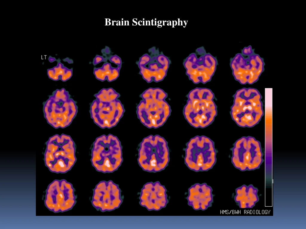
Brain Scintigraphy Normals: Effects of Color Maps
Kansas University Brain Scintigraphy
Brain Scintigraphy Decreased perfusion right side large CVA
Brain Scintigraphy Decreased activity on contralateral cerebellum Cross cerebellar dischisis
Brain Scintigraphy Luxury perfusion Occurs when there is increased blood flow and reduced O2 in brain tissue. Causes acute lactic-metabolic acidosis. Many different disease states can cause this: CVA, trauma, tumor, etc.
Brain Scintigraphy Diamoix HMPAO MRI Harvard Ischemia
Brain Scintigraphy Diamoix HMPAO Ischemia
Brain Scintigraphy Central area of infarction with regional ischemia. Revascularization?
Brain Scintigraphy Central area of infarction with regional ischemia. Revascularization?
Brain Scintigraphy Transverse Slices Tc99m HMPAO Normal
Brain Scintigraphy Transverse Slices Tc99m HMPAO Globally decreased function (particularly pariatal lobes) /decreased gray mater Alzheimer's Disease
Brain Scintigraphy Transverse Slices Tc99m HMPAO Alzheimer's Disease Transverse Slices Tc99m HMPAO Normal
Brain Scintigraphy Coronal Slices Tc99m HMPAO Alzheimer's Disease
Brain Scintigraphy Sagittal Slices Tc99m HMPAO Alzheimer's Disease
Brain Scintigraphy Patchy diffuse areas of increased and decreased activity Differential diagnosis: Multi-infarct Dementia Cocaine Abuse
Brain Scintigraphy Initial scan Diagnosis Cocaine abuse 6 month follow-up
Brain Scintigraphy Brain Multi-infact Transverse
Brain Scintigraphy Increased perfusion to temporal lobe. Herpes Encephalitis
Brain Scintigraphy melenoma MRI.1t2 Tl201
Brain Scintigraphy MRI / Tl201 Overlay Harvard
Brain Scintigraphy Melanoma HMPAO
Brain Scintigraphy MRI Tl201 HMPAO Melanoma
Brain Scintigraphy Melanoma HMPAO MRI Overlay
Brain Scintigraphy Glioblastoma
Brain Scintigraphy Tl Brain SPECT Previous Radiation for Glioblastoma
Brain Scintigraphy 48 hours Mallincrodt Slow movement over convexities Activity in lateral ventricles NPH
Brain Scintigraphy Delayed movement over convexities at 48 hours.
Brain Scintigraphy Delayed movement over convexities at 48 hours. LEAP on posterior
24 hour rt 24 hr ant immediate 48 hr lt 48 hr ant 48 hr rt Brain Scintigraphy Same patient with laterals
Brain Scintigraphy 4 hour 24 hour 48 hour
Brain Scintigraphy Shunt Patency
Brain Scintigraphy Non-patient Shunt At 20 minutes following injection of tracer into the shunt reservoir, activity is seen in the proximal limb tubing only. At 40 minutes, activity is seen in the lateral ventricles, and third and fourth ventricles, as well as extending down the spinal canal. Still, no activity is seen in the distal shunt.
Brain Scintigraphy Occluded Shunt?
Brain Scintigraphy Abdominal Images
Brain Scintigraphy Displaying the initial images at higher intensity allows visualization of tiny lymphatic channels draining activity away from an infiltrated dose
Brain Scintigraphy Flow study
Brain Scintigraphy Flow for brain death
Brain Scintigraphy Is this Brain Death? Return to the Table of Contents