Download

1 / 26
270 likes | 434 Vues
Corneal Iron Ring After Hyperopic LASIK. Grand Rounds April 2005 Jay C. Bradley, MD David L. McCartney, MD. Report of Case:. 56 year old male Pre-operative refraction +6.00 –2.50 x 7 +6.00 –1.75 x 165 Pre-operative pachymetry OD – 516 μ m OS – 525 μ m.

E N D
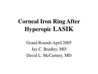
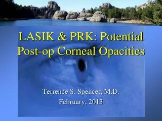
Corneal Iron Ring After Hyperopic LASIK Grand Rounds April 2005 Jay C. Bradley, MD David L. McCartney, MD
Report of Case: • 56 year old male • Pre-operative refraction +6.00 –2.50 x 7 +6.00 –1.75 x 165 • Pre-operative pachymetry OD – 516 μm OS – 525 μm
Operative Summary (2001): • LADARVision 4000 • Laser-assisted in situ keratomileusis • Blend Radius 1.50 mm • Zone Size 6.00 mm • Ablation depth OD – 91.0 microns OS – 90.8 microns • No flap or ablation complications
Follow-up (2004) • Returns to clinic with complaints of decreased near vision OU • Does not use distance Rx • Normal exam except for trace NSC OU and prominent corneal iron ring OU (~ 5 mm diameter) • Patient given Rx for reading glasses
Prior Reports of Corneal Iron Ring After Hyperopic LASIK • Ozdamar A et al. Cornea. 1999 Mar; 18(2):243-5. • 3 patients with pre-op spherical equivalent refraction of +3.37 to +6.50 • Paracentral iron ring OU at 6 – 7 months after surgery • Localized to outside border of central steep zone • No change in ring at 1 year follow-up
Prior Reports of Corneal Iron Ring After Hyperopic LASIK • Probst LE et al. JCRS. 1999 May; 25:868-870. • 1 patient with pre-op Rx of +7.00 –1.50 x 153 OD and +7.50 –1.50 x 165 OS • Paracentral corneal iron ring OU at 6 months after surgery (5 mm diameter) • 6.0 mm optical zone with 9.5 mm blend zone • Ring corresponded to base of hyperopic ablation on corneal topography • Increased mean pre-operative SE and ablation depth associated with deposition
Histology • Hemosiderin deposition in basal corneal epithelial cells • Iron deposits when there is an abrupt change in corneal surface curvature in area of diminished tear flow and hydrodynamic stasis (Hyperopic PRK)
Pathogenesis: Tear-pool hypothesis • Proposed by Gass (1964) • Sequestered iron in the tear film is preferentially deposited in areas of the cornea with pooling of the tear film • Explains Hudson-Stahli lines corresponding to the lid position at rest • Low [iron] in tears, protective effect of mucus, and occurrence of iron lines in eyes without tears question this theory
Pathogenesis: Basal-Cell-Migration Theory • Proposed by Rose and Lavin (1987) • Abrasive lid action on corneal surface induces enhanced mitotic activity • Dividing, nonmigrating, basal cells become mature and accumulate iron
Pathogenesis: Tear Desiccation Hypothesis • Proposed by Assil (1993) • Iron deposits occur in areas of initial tear breakup
Pathogenesis: Senescent Basal Cell Hypothesis • Proposed by Assil (1993) • Iron accumulates in the epithelial cells where there are diminished rates of cell turnover
Iron Lines Associated with Rx / Tx Corneal Procedures • Arcuate along anterior suture border after PK • Lamellar keratoplasty • Ring adjacent to margin of donor lenticule after epikeratophakia • Central stellate pattern after RK (~ 80 %) • Inferior line or midperipheral arcuate after intrastromal corneal ring segment (ICRS) placement • Central line after uneventful PRK (myopic and hyperopic) • Small central ring associated with steep central islands after PRK • Myopic and hyperopic LASIK • Overnight orthokeratology • Conductive keratoplasty
Other Corneal Iron Lines • Fleischer ring • Associated with keratoconus • First reported in 1906 • Partial or complete ring encircling base of cone • Yellowish to dark brown • Best seen with cobalt blue light
Other Corneal Iron Lines • Hudson-Stähli • Iron line along lower third of cornea in otherwise normal eyes • Associated with age • Area of tear pooling when eye lids at rest
Other Corneal Iron Lines • Stocker-Busacca line • Associated with pterygia • Anterior to leading edge • Yellow to golden brown
Other Corneal Iron Lines • Ferry’s line • Associated with filtering blebs • First reported in 1968
Other Corneal Iron Lines • Elevated corneal scars • Salzmann’s nodular degeneration • Corneal foreign body • Coats’ white ring • Juvenile corneal arcus lipoides
Importance of Corneal Iron Lines • Diagnosis of associated ocular condition • Identification of corneal donor tissue with prior refractive surgery (ie LASIK flap) • Possible estimation of optical size and centration of ablation with hyperopic LASIK