Download
1 / 28
300 likes | 876 Vues
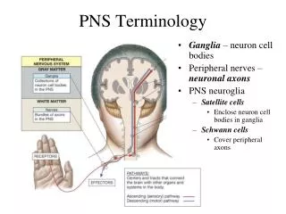
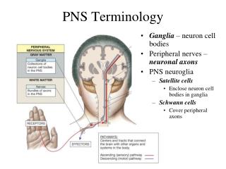
PNS Terminology. Ganglia – neuron cell bodies Peripheral nerves – neuronal axons PNS neuroglia Satellite cells Enclose neuron cell bodies in ganglia Schwann cells Cover peripheral axons. The Cranial Nerves (PNS). I - Olfactory II - Optic III - Oculomotor IV-Trochlear V - Trigeminal

E N D
PNS Terminology • Ganglia – neuron cell bodies • Peripheral nerves – neuronal axons • PNS neuroglia • Satellite cells • Enclose neuron cell bodies in ganglia • Schwann cells • Cover peripheral axons
I - Olfactory II - Optic III - Oculomotor IV-Trochlear V - Trigeminal VI - Abducens VII - Facial VIII – Acoustic/Vestibulocochlear IX - Glossopharyngeal X - Vagus XI – Accessory/Spinal Accessory XII - Hypoglossal -cranial nerves – 12 pairs -considered part of the peripheral nervous system (PNS) -olfactory & optic & acoustic contain only sensory axons = sensory nerves -some carry motor information – motor nerves e.g. oculomotor, trochlear, abducens -remaining are mixed nerves – both motor and sensory axons “some say my mother bought my brother some bitter beer – my, my”
I III II V VI VII IX VIII X Optic Chiasma
The Olfactory Nerve (I) • Carries sensory information • Sense of smell • Synapse within olfactory bulbs
The optic nerve (II) • Carries visual information
The oculomotor nerve (III) • Primary source of innervation for extra-ocular muscles • Move the eyeball • The trochlear nerve (IV) • Smallest cranial nerve • Innervates superior oblique eye muscle • The abducens nerve (VI) • Innervates lateral rectus muscle of eye
The Trigeminal Nerve (V) • Largest cranial nerve • Mixed nerve • sensory – touch, pain & thermal • Ophthalmic branch • sensory – upper eyelid, eyeball lacrimal glands, side of nose, forehead and scalp • Maxillary branch • sensory – nose, palate, part of pharynx, upper teeth, upper lip and lower eyelid • Mandibular branch • sensory – tongue, cheek, lower teeth, skin over mandible and side of head anterior to ear -motor – muscles of chewing
The Facial Nerve (VII) • Mixed nerve • Controls muscles of scalp and face • Pressure sensations from face • Taste sensations from tongue
The Vestibulocochlear Nerve (VIII) • Vestibular nerve • Monitors sense of balance, position and movement • Cochlear nerve • Monitors hearing
The Glossopharyngeal Nerve (IX) • Mixed nerve • Innervates the tongue • Controls swallowing
The Vagus Nerve (NX) • Mixed nerve • Vital to autonomic control of visceral function
The accessory nerve (XI) • Internal branch • Innervates swallowing muscles • External branch • Controls muscles associated with pectoral girdle • The hypoglossal nerve (XII) • Voluntary motor control over tongue movements
31 Pairs of Spinal Nerves • Ensheathed by three connective tissue layers • Outermost epineurium • Dense network of collagen fibers • Middle perineurium • Partitions nerve into fascicles • Inner endoneurium • Delicate connective tissue fibers surrounding each axon • Under the endoneurium is the myelin sheath – outer layer is called the neurilemma • Neurilemma covers the myelin sheath and Schwann cells • Myelin sheath covers the axon
Spinal Nerves • connected to the spinal cord via roots (bundles of axons) • Posterior root = sensory axons into the posterior gray horn • Anterior root = motor axons from the anterior gray horn • before the posterior root is the dorsal root ganglion - cell bodies of incoming sensory neurons (axons continue on to form the root) • emerge from intervertebral foramina as mixed nerves
Spinal Nerve • after passing through intervertebral foramina the spinal nerve branches into three rami • Dorsal ramus • -sensory/motor innervation to skin and muscles of back • Ventral ramus • - Sensory/motor innervation to ventral and lateral body surface/skin, body wall structures, muscles of the upper and lower limbs
rami communicantes = • Third branch from the spinal nerve • -carries nerves of the ANS
Dorsal Root of SN Ventral Root of SN SPINAL NERVE Dorsal Ramus Ventral Ramus Rami Communicantes Sensory – IN Motor – OUT SKIN BACK MUSCLES Sensory – IN Motor – OUT TRUNK LIMBs Signals to and from the ANS VISCERA – cardiac and Smooth muscle
Epidural space Dura and Arachnoid maters Dorsal Ramus Dorsal Root Ventral Ramus Dorsal Root Ganglion Ventral Root Rami Communicantes
Nerve Plexuses • Joining of ventral rami of spinal nerves to form nerve networks or plexuses • Found in neck, arm, low back & sacral regions • No plexus in thoracic region • intercostal nn. innervate intercostal spaces • T7 to T12 supply abdominal wall as well • Four major plexuses • Cervical plexus • Brachial plexus • Lumbar plexus • Sacral plexus
Cervical Plexus • Cervical plexus • C1-C4 ventral rami • Some fibers from C5 • Innervates muscles of the neck and diaphragm • Phrenic nerve
Brachial Plexus • Ventral rami of C5-T1 • Innervates pectoral girdle and upper limbs • Nerves arise from cords or trunks • Superior, middle and inferior trunks • Lateral, medial and posterior cords • Superior and Middle trunk contribute to the Lateral cord (“SML”) -Superior, middle and inferior trunk all contribute to the Posterior cord (“SMIP”) -inferior trunk continues on as the Medial cord (“IM”)
Lumbar and Sacral Plexuses • Lumbar plexus - ventral rami of T12–L4 • Sacral plexus – ventral rami of L4–S4 • Innervate pelvic girdle and lower limbs
External Anatomy of Spinal Cord Some nerves to know -phrenic -ulnar -radial -medial -musculocutaneous -femoral -obturator -sciatic -ilioinguinal -thoracic (intercostals)