Download
1 / 34
340 likes | 371 Vues
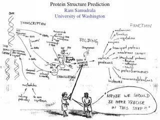
This lecture covers the basics of protein structure, including the primary structure, amino acid anatomy, R-group characteristics, peptide bonds, secondary structure (alpha-helix, beta-sheet, and turns), tertiary and quaternary structures, and various classes of proteins.

E N D
Primary structure = order of amino acids in the protein chain
Charged/polar R-groups generally map to surfaces on soluble proteins
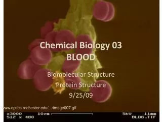
Non-polar R-groups tend to be buried in the cores of soluble proteins Myoglobin Blue = non-polar R-group Red = Heme
protonated unprotonated ( ) Some R-groups can be ionized The Henderson-Hasselbalch equation allows calculation of the ratio of a weak acid and its conjugate base at any pH
General protein pK’ values • Approximate pK' • Group In a “Typical” Protein • -carboxyl (free) 3 (C-terminal only) • -carboxyl (Asp) 4 • -carboxyl (Glu) 4 • imidazole (His) 6 • sulfhydryl (Cys) 8 • 1˚-amino (free) 8 (N-terminal only) • -amino (Lys) 10 • hydroxyl (Tyr) 10 • 2˚-amino (Pro)(free) 9 (N-terminal only) • guanido (Arg) 12
An example of a Henderson-Hasselbalch calculation • What is the structure of the histidine side chain at pH 4? 4 = 6.0 - log [HB]/[B-] -2 = -log [HB]/[B-] 2 = log [HB]/[B-] 100 = [HB]/[B-] • So, in a solution of histidine at pH 4, the majority structure is that of the protonated form.
Amino Acids Are Joined By Peptide Bonds In Peptides - a-carboxyl of one amino acid is joined to a-amino of a second amino acid (with removal of water) - only a-carboxyl and a-amino groups are used, not R-group carboxyl or amino groups
The peptide bond is planar This resonance restricts the number of conformations in proteins -- main chain rotations are restricted tof and y.
small hydrophobic large hydrophobic polar positive charge negative charge DnaG E. coli ...EPNRLLVVEGYMDVVAL... DnaG S. typ ...EPQRLLVVEGYMDVVAL... DnaG B. subt ...KQERAVLFEGFADVYTA... gp4 T3 ...GGKKIVVTEGEIDMLTV... gp4 T7 ...GGKKIVVTEGEIDALTV... : : * * * : : : : Primary sequence reveals important clues about a protein • Evolution conserves amino acids that are important to protein structure and function across species. Sequence comparison of multiple “homologs” of a particular protein reveals highly conserved regions that are important for function. • Clusters of conserved residues are called “motifs” -- motifs carry out a particular function or form a particular structure that is important for the conserved protein. motif
Generally only a limited amount of a protein’s surface is well conserved Invariant (the residue is always the same, e.g. Asp) Conserved (the residue is generally similar, e.g. negatively charged) Not conserved (can be many different residues in different species)
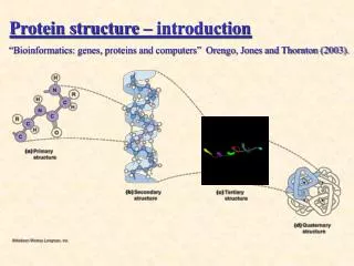
Secondary structure = local folding of residues into regular patterns
The a-helix • In the a-helix, the carbonyl oxygen of residue “i” forms a hydrogen bond with the amide of residue “i+4”. • Although each hydrogen bond is relatively weak in isolation, the sum of the hydrogen bonds in a helix makes it quite stable. • The propensity of a peptide for forming an a-helix also depends on its sequence.
The b-sheet • In a b-sheet, carbonyl oxygens and amides form hydrogen bonds. • These secondary structures can be either antiparallel (as shown) or parallel and need not be planar (as shown) but can be twisted. • The propensity of a peptide for forming b-sheet also depends on its sequence.
b turns • b-turns allow the protein backbone to make abrupt turns. • Again, the propensity of a peptide for forming b-turns depends on its sequence.
Which residues are common for a-helix, b-sheet, and b-turn elements?
Ramachandran plot -- showsf and yangles for secondary structures
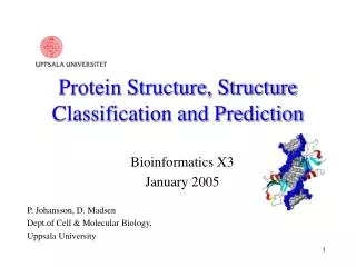
Example of tertiary and quaternary structure - PriB homodimer Example is PriB replication protein solved at UW: Lopper, Holton, and Keck (2004) Structure12, 1967-75.
Example of quaternary structure - Sir1/Orc1 heterodimer Example is Sir1/Orc1 complex solved at UW: Hou, Bernstein, Fox, and Keck (2005) Proc. Natl. Acad. Sci.102, 8489-94.
Examples of other quaternary structures TetramerHexamerFilament SSBDNA helicaseRecombinase Allows coordinated Allows coordinated DNA binding Allows complete DNA binding and ATP hydrolysis coverage of an extended molecule
Classes of proteins Functional definition: Enzymes: Accelerate biochemical reactions Structural: Form biological structures Transport: Carry biochemically important substances Defense: Protect the body from foreign invaders Structural definition: Globular: Complex folds, irregularly shaped tertiary structures Fibrous: Extended, simple folds -- generally structural proteins Cellular localization definition: Membrane: In direct physical contact with a membrane; generally water insoluble. Soluble: Water soluble; can be anywhere in the cell.