The Muscular System
960 likes | 998 Vues
The Muscular System. Function of Muscles. Muscles are responsible for four functions Produce movement Contraction! Skeletal : Run away from danger, smile in flirtation Smooth : peristalsis of digestion Cardiac : beat of heart Maintain posture

The Muscular System
E N D
Presentation Transcript
Function of Muscles • Muscles are responsible for four functions • Produce movement • Contraction! • Skeletal: Run away from danger, smile in flirtation • Smooth: peristalsis of digestion • Cardiac: beat of heart • Maintain posture • Upright/seated – fight downward pull of gravity
Stabilize joints • Tendons attaching muscles to bones of joints for stabilization
Generate heat • By-product of muscle activity produces heat • As ATP (adenosine triphosphate) is used to power contraction, ¾ energy released as heat • Maintains normal body temperature (homeostasis!) • Skeletal muscle (40% body mass) most responsible
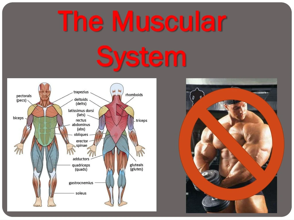
Types of Muscles • Three basic muscle types are found in the body • Skeletal muscle - attached to bones • Smooth muscle - hollow visceral (internal) organs • Cardiac muscle – heart
Skeletal Muscles • Most are attached by tendons to bones • Cells are multinucleate (many nuclei in one cell) • Striated – have visible banding • Voluntary – subject to conscious control • Cells are surrounded and bundled by connective tissue Vertical striations (up & down)
“Packaging” of Skeletal Muscles • Muscle fiber = muscle cell • Endomysium – around single muscle fiber • Fascicle = bundle of fibers • Perimysium – around a fascicle • Typical skeletal muscle = bundle of fascicles • Epimysium – covers the entire skeletal muscle • Fascia – on the outside of the epimysium Figure 6.1
Epimysium blends into a connective tissue attachment • Tendon – cord-like structure • Aponeuroses – sheet-like structure • Sites of muscle attachment • Bones • Cartilages • Connective tissue coverings
Smooth Muscle • Has no striations • Spindle-shaped cells • Single nucleus • Involuntary – no conscious control • Found mainly in the walls of hollow organs Figure 6.2a
Cardiac Muscle • Has striations • Usually has a single nucleus • Joined to another muscle cell at an intercalated disc • Involuntary • Found only in the heart Figure 6.2b
Anatomy of Skeletal Muscle • Cells are multinucleate (many nuclei per cell) • Nuclei (oval-shaped) are just beneath the sarcolemma (specialized plasma membrane) • Contain sarcoplasmic reticulum (specialized smooth endoplasmic reticulum [ER]) for Ca2+ storage Figure 6.3a
One muscle fiber (muscle cell) is made of a bundle of myofibrils (organelles) that nearly fill cytoplasm Figure 6.3a
One myofibril (organelle) is made of chains of tiny contractile units called sarcomeres (Z disc to Z disc) SARCOMERE
one sarcomere is made of two myofilaments (protein filaments): • actin (thin) and myosin (thick)
Thick filaments • Composed of the protein myosin • Has ATPase enzymes which break ATP bonds to make energy for muscle contraction! • Thin filaments • Composed of the protein actin Figure 6.3c
Myosin filaments have heads (extensions, or cross bridges which look like golf club heads) that move • Myosin and actin overlap somewhat Figure 6.3d
RECAP • Muscle fiber • Myofibrils • Sarcomere • Myofilaments • Actin • Myosin Site of muscle contraction
Alignment of myofibrils in sarcomere creates banding pattern • I band (light band) = absence of myosin • A band (dark band) = concentration of myosin Figure 6.3b
Z disc =Area where actin joins other actin molecules, forms Z-shaped pattern • H zone (bare zone) = area that lacks thin filaments • M line = center of H zone that contains tiny protein rods that hold adjacent myosin proteins together
Banding pattern changes shape during contraction when each sarcomere segment gets shortened (H zone gets smaller)
Muscle Activity: Contraction • Muscle has two functional properties that are necessary for contraction: • Irritability – ability to receiveand respondto a stimulus (nerve to muscle) • Contractility – ability to shortenwhen an adequate stimulus is received via sliding filament theory (inside muscle)
Irritation: Nerve Stimulus to Muscles • Skeletal muscles must be stimulated by a nerve • Motor units are responsible for stimulating a few to hundreds of muscle cells. Consist of • One neuron • All muscle cells stimulated by that one neuron Figure 6.4a
Neuron (nerve cell) relays message through electrical and chemical signals. Consists of • Cell body (with dendrite fingers) • Axon or nerve fiber (with axon terminal projections)
Axon terminals approach (never physically touch) muscle’s sarcolemma via neuromuscular junctions • Gap between axon terminals and muscle cells called synaptic cleft • Filled with interstitial (“between tissues”) fluid Figure 6.5b
Steps of Nerve Stimulation (BEFORE MUSCLE) • Electrical nerve impulse reaches dendrites
Signal moves down cell body, to axon terminals, signals vesicles with chemical neurotransmitter called acetylcholine (ACh) to move to nerve membrane
ACh is released via exocytosis into synaptic cleft (space between nerve and muscle) • ACh attaches to receptors on the sarcolemma membrane • Triggers Na+/K+ (sodium/potassium) pumps to open, making sarcolemma permeable to Na+ (Na+ rushes in, K+ rushes out)
Rapid change in ion movement (sodium in, potassium out of the cell) is called depolarization which generates an electrical current called action potential • Action potential will start, muscle contraction which cannot be stopped travels over entire surface of sarcolemma
Action Potential Analogy Polar = opposite ends INSIDERS outsiders outsiders outsiders outsiders INSIDERS INSIDERS INSIDERS INSIDERS INSIDERS INSIDERS outsiders outsiders INSIDERS outsiders outsiders Polarized (rest) Depolarized (action potential) Repolarized (opposite poles again but switched sides) Polarized
In the meantime, ACh is now broken down by enzyme (AChase) to acetic acid and choline • This is why one nerve impulse produces one contraction, otherwise one nerve impulse would cause continual contraction & we’d run out of energy!
RECAP - Steps of Nerve Stimulation • Nerve cell receives electrical stimulus through dendrites • Stimulus send down cell body, out through axon terminals as chemical signal • ACh neurotransmitter released from vesicles into synaptic cleft • Receptors on sarcolemma receive ACh • Sarcolemma now permeable to Na+ ions = Na+ rushes in, K+ rushes out • Depolarization of muscle cells creates action potential
2. Contraction at the Muscle: The Sliding Filament Theory • Activation by nerve causes myosin heads (cross bridges) to attach to binding sites on the thin (actin) filament • Myosin heads then bind to the next site of the thin filament due to Ca2+ and ATP Figure 6.7
Ca2+ from sarcoplasmic reticulum which was triggered to release Ca2+ as a result of action potential • This continued action causes a sliding of the myosin along the actin • The result is that the muscle is shortened (contracted) Figure 6.7
Muscle cell relaxes again when: • 1. diffusion of K+ ions out of cell • 2. Na+/K+ pump brings ions back to original state (Na+ back out, K+ back in)
Action Potential Analogy Polar = opposite ends INSIDERS outsiders outsiders outsiders outsiders INSIDERS INSIDERS INSIDERS INSIDERS INSIDERS INSIDERS outsiders outsiders INSIDERS outsiders outsiders Polarized (rest) Depolarized (action potential) Repolarized (opposite poles again but switched sides) Polarized
Recap - Sliding Filament Contraction • Action potential triggers sarcoplasmic reticulum to release Ca2+ • Action potential causes myosin heads to attach to binding sites on actin filament • Ca2+ with ATP triggers myosin heads to attach to neighbor binding sites on same actin filament, causing actin to move toward each other = contraction!
Contraction Responses • Muscle fiber contraction is “all or none” • Within a skeletal muscle, not all fibers may be stimulated during the same interval • Different combinations of muscle fiber contractions may give differing responses • Graded responses – different degrees of skeletal muscle shortening
Types of Graded Responses • Twitch • Single, brief contraction • Not a normal muscle function Figure 6.9a–b
Tetanus (summing of contractions) • One contraction is immediately followed by another • The muscle does not completely return to a resting state • The effects are added (summed) Figure 6.9a–b
Fused (complete) tetanus • No evidence of relaxation before the following contractions • The result is one sustained muscle contraction Figure 6.9c–d


