Download
1 / 1
10 likes | 142 Vues
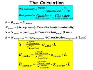
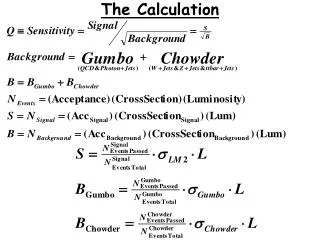
Post-Processing of the Finite Element Model of a Cornea for Collagen Crosslinking Treatment of Keratoconus Daniel H. Kang , RET Fellow 2011 Lindblom Math and Science Academy RET Mentor: Dr. Craig Foster, PhD NSF- RET Program. Introduction. Background.

E N D
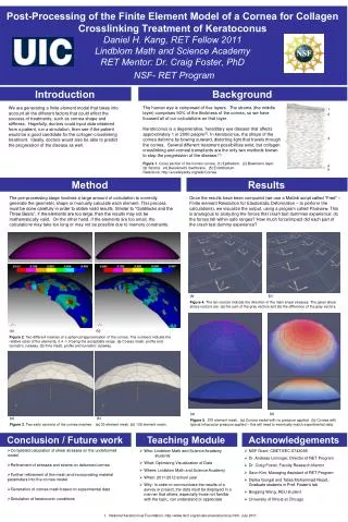
Post-Processing of the Finite Element Model of a Cornea for Collagen Crosslinking Treatment of KeratoconusDaniel H. Kang, RET Fellow 2011Lindblom Math and Science AcademyRET Mentor: Dr. Craig Foster, PhDNSF- RET Program Introduction Background The human eye is composed of five layers. The stroma(the middle layer) comprises 90% of the thickness of the cornea, so we have focused all of our calculations on that layer. Keratoconus is a degenerative, hereditary eye disease that affects approximately 1 in 2000 people[1]. In keratoconus, the shape of the cornea deforms by bowing outward, distorting light that travels through the cornea. Several different treatment possibilities exist, but collagen crosslinking and corneal transplants are the only two methods known to stop the progression of the disease.[1] We are generating a finite element model that takes into account all the different factors that could effect the success of treatments, such as cornea shape and stiffness. Hopefully, doctors could input data obtained from a patient, run a simulation, then see if the patient would be a good candidate for the collagen crosslinking treatment. Ideally, doctors would also be able to predict the progression of the disease as well. 1 2 3 Figure 1. Cross section of the human cornea. (1) Epithelium. (2) Bowman’s layer. (3) Stroma. (4) Descemet’s membrane. (5) Endothelium. Reference: http://en.wikipedia.org/wiki/Cornea 4 5 Method Results The pre-processing stage involves a large amount of calculation to correctly generate the geometric shape or manually calculate each element. This process must be done carefully in order to obtain valid results. Similar to “Goldilocks and the Three Bears”, if the elements are too large, then the results may not be mathematically valid. On the other hand, if the elements are too small, the calculations may take too long or may not be possible due to memory constraints. Once the results have been computed (we use a Matlab script called “Fred” – Finite element Resolution for Elastostatic Deformation – to perform the calculations), we visualize the output, using a program called Paraview. This is analogous to analyzing the forces that crash test dummies experience: do the forces fall within safe ranges? How much force/impact did each part of the crash test dummy experience? (a) (b) Figure 4.The tan vectors indicate the direction of the main shear stresses. The given shear stress vectors are: (a) the sum of the gray vectors and (b) the difference of the gray vectors. (a) (b) Figure 2. Two different meshes of a spherical approximation of the cornea. The numbers indicate the relative sizes of the elements, 0.4 -1.0 being the acceptable range. (a) Coarse mesh, profile and isometric cutaway. (b) Fine mesh, profile and isometric cutaway. (a) (b) Figure 5. 570 element mesh. (a) Cornea model with no pressure applied. (b) Cornea with typical intraocular pressure applied – this will need to eventually match experimental data. (a) (b) Figure 3.Two early versions of the cornea meshes. (a) 30 element mesh. (b) 108 element mesh. Conclusion / Future work Teaching Module Plan Acknowledgements • Who: Lindblom Math and Science Academy students • What: Optimizing Visualization of Data • Where: Lindblom Math and Science Academy • When: 2011-2012 school year • Why: In order to communicate the results of a survey or project, the data must be displayed in a manner that others, especially those not familiar with the topic, can understand or appreciate. • NSF Grant: CBET-EEC-0743068 • Dr. Andreas Linninger, Director of RET Program • Dr. Craig Foster, Faculty Research Mentor • Seon Kim, Managing Assistant of RET Program • DipikaGongaland Talisa Mohammad Nejad, Graduate students in Prof. Foster’s lab • Bingqing Wang, REU student • University of Illinois at Chicago • Completed calculation of shear stresses on the undeformed model. • Refinement of stresses and strains on deformed cornea • Further refinement of the mesh and incorporating material parameters into the cornea model • Generation of cornea mesh based on experimental data • Simulation of keratoconic conditions • National Keratoconus Foundation. http://www.nkcf.org/en/about-keratoconus.html. July 2011.