
The Brain
The Brain. Cerebral Hemispheres. How is the brain organized? Define the major lobes Distinguish between gyri and sulci Compare the functional areas of the cerebral cortex. The cerebrum is divided into three major areas: Cerebral Cortex Internal White Matter Basal Nuclei.

The Brain
E N D
Presentation Transcript
Cerebral Hemispheres How is the brain organized? • Define the major lobes • Distinguish between gyri and sulci • Compare the functional areas of the cerebral cortex
The cerebrum is divided into three major areas: • Cerebral Cortex • Internal White Matter • Basal Nuclei
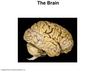
The cerebral hemispheres form the superior part of the brain. It makes up most of the mass of the brain.
The Cerebral Cortex The cortex is composed of gray matter which contains neuron cell bodies, dendrites, glial cells and blood vessels. It is only 2 to 4 mm in thickness. The folds increase the surface area.
The surface of the hemispheres is marked by elevated ridges called gyri and shallow grooves called sulci.
Lissencephaly, which literally means smooth brain, is a rare brain formation disorder caused by defective neuronal migration during the 12th to 24th weeks of gestation resulting in a lack of development of brain folds (gyri) and grooves (sulci).
Normal Brain Lissencephaly
The prognosis for children with lissencephaly varies depending on the malformation. Many individuals remain in a 3-5 month developmental level, while others may appear to have near normal intelligence and development. Some children with lissencephaly will be able to roll over, sit, reach for objects, and smile socially.
The cortex contains three kinds of functional areas: • Motor area • Sensory areas • Association areas
Each hemisphere is primarily concerned with sensory and motor function on the opposite (contralateral) side of the body.
Each hemisphere is not equal in function. There is lateralization (specialization) of cortical functions. Left hemisphere is logical while the right is creative
The motor areas control voluntary movement and are found primarily in the posterior part of the frontal lobes and include: • the primary motor cortex, • premotor cortex, • Broca’s area and • the frontal eye field.
Figure 12.8a Functional and structural areas of the cerebral cortex. Motor areas Sensory areas and related association areas Central sulcus Primary motor cortex Primary somatosensory cortex Premotor cortex Somatic sensation Frontal eye field Somatosensory association cortex Broca’s area (outlined by dashes) Gustatory cortex (in insula) Taste Prefrontal cortex Wernicke’s area (outlined by dashes) Working memory for spatial tasks Executive area for task management Primary visual cortex Working memory for object-recall tasks Vision Visual association area Solving complex, multitask problems Auditory association area Hearing Primary auditory cortex (a) Lateral view, left cerebral hemisphere Motor association cortex Primary sensory cortex Primary motor cortex Sensory association cortex Multimodal association cortex
Primary motor cortex is located in the precentral gyrus of the frontal lobe. Their function is to control the precise voluntary movements of the skeletal muscles.
Figure 12.9 Body maps in the primary motor cortex and somatosensory cortex of the cerebrum. Posterior Motor Sensory Anterior Motor map in precentral gyrus Sensory map in postcentral gyrus Toes Genitals Jaw Primary motor cortex (precentral gyrus) Primary somato- sensory cortex (postcentral gyrus) Tongue Intra- abdominal Swallowing
Common disorders include stroke, tumors or traumatic brain injury. Effects include loss of specific motor functions. Stroke
Premotor cortex is located just anterior precentral gyrus of the frontal lobe. It's function is to control learned motor skills of a repetitive or patterned nature, for example typing.
Disorders of the premotor cortex are commonly due to stroke, tumors or trauma. One condition is known as Apraxia or the inability to recognize objects by touch.
Broca’s Area lies anterior and inferior to the premotor cortex. It is typically found only in the left hemisphere and is the motor speech area.
Aphasia is a disorder caused by damage to the parts of the brain that control language. It can make it hard for you to read, write, and say what you mean to say.
There are four main types: • Expressive aphasia - you know what you want to say, but you have trouble saying or writing what you mean
There are four main types: • Expressive aphasia • Receptive aphasia - you hear the voice or see the print, but you can't make sense of the words
There are four main types: • Expressive aphasia • Receptive aphasia • Anomic aphasia - you have trouble using the correct word for objects, places, or events
There are four main types: • Expressive aphasia • Receptive aphasia • Anomic aphasia • Global aphasia - you can't speak, understand speech, read, or write Aphasia
Frontal eye field is located on or near the premotor cortex and is above Broca’s area. This region controls the voluntary movement of the eyes.
Thesensory area of the cerebral cortex is concerned with conscious awareness. Areas involved with this function are found in the parietal, temporal and occipital lobes.
Primary somatosensory cortex receive information from the sensory receptors in the skin and proprioceptors (position receptors) in the skeletal muscles, joints and tendons.
Figure 12.9 Body maps in the primary motor cortex and somatosensory cortex of the cerebrum. Posterior Motor Sensory Anterior Motor map in precentral gyrus Sensory map in postcentral gyrus Toes Genitals Jaw Primary motor cortex (precentral gyrus) Primary somato- sensory cortex (postcentral gyrus) Tongue Intra- abdominal Swallowing
Somatosensory association cortex serves to integrate sensory information such as temperature, pressure to produce an understanding of what is being touched.
Association Areas Multimodal association areas receive input from multiple senses and sends outputs to multiple areas. There are 3 main areas: • Anterior association area • Posterior association area • Limbic association area
Anterior association area Is located in the frontal lobe. It is considered the most complicated and is involved with learning (cognition), recall and personality. Additional functions include working memory, abstraction and planning.
Posterior association area Covers the temporal, parietal and occipital lobes. This area is involved with pattern recognition. It is what allows us to recognize familiar places. An additional function understands of written and spoken language.
Isolated problems with these association areas are rare. Trauma or stroke often leads to large areas of affliction. The case of Phineas Gage
Limbic association area This area is sometimes called the pleasure center. In animal studies it is correlated with sexual arousal, behavior and olfaction (think about this last one). Big Bang
Cerebral White Matter The cerebral white matter is responsible for communicating between the cerebral areas and the lower CNS centers. This area consists largely of myelinated fibers bundled into large tracts.
The white fibers or tracts are classified based on which direction they run. • Commissural fibers connect gray areas of the two hemispheres allowing them to coordinate. • Association fibers connect different parts of the same hemisphere • Projection fibers either enter the cerebral cortex from the lower brain or descend to the lower areas from the cortex.
Figure 12.10 Types of fiber tracts in white matter. Association fibers Longitudinal fissure Superior Commissural fibers (corpus callosum) Lateral ventricle Corona radiata Basal nuclei • Caudate • Putamen • Globuspallidus Fornix Internal capsule Thalamus Gray matter Third ventricle White matter Projection fibers Pons Decussation of pyramids Medulla oblongata (a) (b)
Basal Nuclei These represent the third region of the cerebral hemisphere. The basal nuclei consist of the: • caudate nucleus, • putamen and • the globus pallidus.
Together the putamen and globuspallidus form the lentiform nucleus that flanks the internal capsule
Figure 12.11b Basal nuclei (1 of 2). Anterior Cerebral cortex Cerebral white matter Corpus callosum Anterior horn of lateral ventricle Caudate nucleus Putamen Lentiform nucleus Globus pallidus Thalamus Tail of caudate nucleus Third ventricle Inferior horn of lateral ventricle (b) Posterior
Figure 12.11b Basal nuclei (2 of 2). Cerebral cortex Cerebral white matter Corpus callosum Anterior horn of lateral ventricle Caudate nucleus Lentiform nucleus Thalamus Third ventricle Inferior horn of lateral ventricle (b)
The basal nuclei receive input from the entire cerebral cortex. They appear to be important in starting and stopping the intensity of movements initiated by the cerebral cortex. They are important in our ability to multitask.
Disorders of the Basal Nucleus The basal ganglia play a central role in a number of neurological conditions. The most notable are, Parkinson’s disease and Huntington's disease. Basal ganglia dysfunction is also implicated in some other disorders of behavior control such as Tourette’s syndrome, and obsessive compulsive disorder (OCD),


