Chapter 15 Cytoskeleton: Regulation by Accessory Proteins
190 likes | 476 Vues
Chapter 15 Cytoskeleton: Regulation by Accessory Proteins. Microtubules are nucleated by a protein complex containing g -tubulin. Dynamics of cytoskeletal filaments: Length Stability Number Geometry. g- tubulin ring complex ( g -TuRC): minus end. Subcomplex: accessory proteins

Chapter 15 Cytoskeleton: Regulation by Accessory Proteins
E N D
Presentation Transcript
Chapter 15 Cytoskeleton: Regulation by Accessory Proteins
Microtubules are nucleated by a protein complex containing g-tubulin Dynamics of cytoskeletal filaments: Length Stability Number Geometry g-tubulin ring complex (g-TuRC): minus end Subcomplex: accessory proteins bound to g-tubulins In a living cell, cytoskeletal filaments are more dynamic: accessory proteins! By Covalent modification of subunits& Accessory proteins
Microtubule-organizing center (MTOC) Centrisomes star-like, “astral” 2 Centrioles (L-shaped) Centrosome matrix (50 g-TuRCs) Become basal bodies, mitotic spindles Fungi (spindle pole body) and plants (near nuclear envelope) don’t have centrioles but use g-tubulin to nucleate
Actin filaments are often nucleated at the plasma membrane “Cell cortex”: shape and movement of the cell surface Spiky bundles: microvilli or filopodia Protrusive veils: lamellipodia Two Actin-related proteins, ARPs, nucleate actin filaments 45% identical to actin The ARP complex (Arp2/3 complex): minus end Attach to the side of another actin filament:branches Grow new filaments!
Profilin promotes polymerization Thymosin keeps actins in monomer form 50% is soluble 50% in filament Associates with plus end! Plus end
One stathmin binds to two tubulin heterodimers and prevents polymerization Phosphorylation of stathmin inhibits stathmin and increases the rate of elongation and suppress dynamic instability
MAPs (bind along the sides of MTs) in axon and dendrites Of a neuron (MAP2 orange) (unphosporylated tau green) Stablize MT against disassembly
Tropomyosin stabalizes actin filaments Cofilin destablizes actin filaments by twisting the actin Filaments a little more tightly--mechanical stress weakens The contacts between actin subunits:brittle and ADP actin Dissociate from minus end easier. Shorter lived than in test tube. Aka actin depolymerization factor Rapid turn over: Binds to ADP actin
Capping proteins change filament dynamics by binding to ends End-binding proteins can affect dynamics at low [ ] Inactivate plus end Minus end either capped by ARP2/3 or uncapped CapZ (muscle Z band) Stable actin filament in muscle: Plus end-CapZ Minus end-tropomodulin(only binds when coated by tropomyosin) Inositol phospholipid, PIP2, uncaps plus ends
Proteins that bind to microtubule ends Dynamic instability Catastrophins: kinesin-related proteins Capping proteins Also control Positioning: Minus end--g-TuRC Plus end--EB1, which binds to Kar9 Directs spindle MTs into the growing bud during mitosis
Actin filament organizations Organization of MT by Stabilizing proteins: MTOC and MAPs Actin organizing is mediated by a separate class of Proteins (crosslinking) Actin bundles Weblike Bundling proteins Gel-forming proteins
(RBC) Four actin-crosslinking proteins Web-like:spectrin & filamin Bundles! 3D gels! Stress fiber Parallel filaments Actin bundling proteins Focal contacts
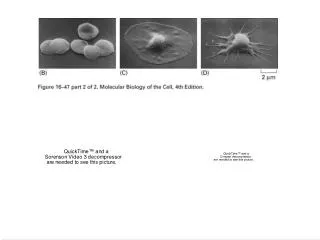
Filamin cross-links actin filaments to a 3-D network Pigment-cell cancers Melanoma cells that have low levels of filamin: blebbing can’t metastasize Lamellipodia is normal and highly metastatic when filamin is restored lamellipodia
Severing proteins regulate the length and kinetic behavior The number of ends increases when severed Faster increase and Decreast of filament mass Gelsolin superfamily Serves at high Ca++. No energy requirement katanin (sword) breaks 13 protofilaments Requires ATP
ERM family (ezrin, radixin and moesin) attaches actin filaments to the plasma membrane Regulated by intracellular and extracellular signals
Focal contacts and stress fibers in a cultured fibroblast Attachments between the actin filaments and ECM that allows cells to pull on the substratum to which they are bound Also relays signals: focal adhesion kinase (FAK)
The dramatic effects of Rac, Rho and Cdc42 on actin organization in fibroblasts Stress fibers Focal contacts Lamellipodia Membrane ruffles Filapodia Microspikes “cytoskeleton?”
Summary Nucleation, polymerization Proteins that bind to the sides End binding proteins Crossing-linking proteins Severing proteins Anchoring to the membrane Rho-family GTPases and regulation by signals