Download
1 / 16
160 likes | 842 Vues
Microscopes . Contents. History Structure Uses Images. History. Invented during the Renaissance

E N D
Contents • History • Structure • Uses • Images
History • Invented during the Renaissance • Lenses had been used to magnify images during the first century A.D., but it was not until the late 1500 hundreds when two Dutch spectacle makers started experimenting with several lenses in a long tube.
The Father of Microscopy • Anton van Leeuwenhoek (1632 – 1723) • he taught himself new methods for grinding and polishing tiny lenses of large curvature • these magnifications of up to 270 diameters • he was the first to see and describe bacteria, yeast plants and the circulation of blood corpuscles in capillaries.
Why do we need microscopes? • Not everything we want to see is visible to the naked eye. • Microscopes uses lenses to brighten and enlarge nearby and very small objects.
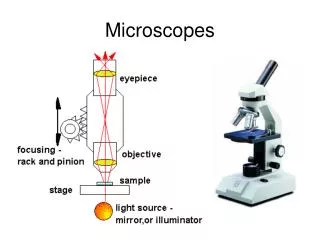
Structure • Microscopes are almost identical to Keplerian telescopes, except for their objective lenses. • Microscopes uses a short focal length objective lens to project a large real image of a nearby object • They must gather large amounts of light from a small object using an Objective Lens
The Objective Lens • The microscope objective lens must be very small and spherical, with a short focal length. • It must be able to: • move in close to the object • catch its rapidly diverging light rays • make those rays converge together as a real image far from the lens • the number marked on the side of the objective lens will tell you how much the image is magnified (for example: x40)
The Objective Lens • The Objective lens is usually a compound lens, or a combination of two lenses made from different types of glass. • The second lens is really there to correct any distortions caused by the first lens, such as chromatic abberation.
The Eyepiece • The eyepiece is the second lens in a microscope. • It is used to magnify the object so it easy to view with your eyes. http://www.physics.carleton.ca/~watson/1000_level/Waves_and_Optics/Gifs/microscope_0.gif
Illumination • Illumination is very important in a microscope. • You can shine a light upward THROUGH the object – which lets you see how much light is absorbed in different places. • You can shine a light downward ONTO the object – which lets you see how light reflects from the surface.
There are four adjustments to be made when using a microscope. • Brightness – adjusts how light or dark the image is using the illumination. • Focus – adjusts if the image is blurry or well defined by using the focus knobs, which change the location of the focal point. • Resolution – defines how easily you can define two adjacent objects from each other and is adjusted by the aperture of the objective lens • Contrast – which is the difference in lighting between two areas. This can be adjusted by changing the intensity of the light and the pinhole aperature.
Uses • Microscopes have been and are being used in many different areas, such as: • identifying minerals • solving crimes • see how freezing affects food • study human cells • find the cause of diseases • discovering where illegal drugs are grown
A hydra getting ready for reproduction http://www.microscope-microscope.org/microscope-images.htm
A paramecium http://www.microscope-microscope.org/gallery/Mark-Simmons/pages/paramecium2.htm
A green protist http://www.microscope-microscope.org/gallery/Mark-Simmons/pages/the_fish.htm