Download
1 / 34
380 likes | 874 Vues
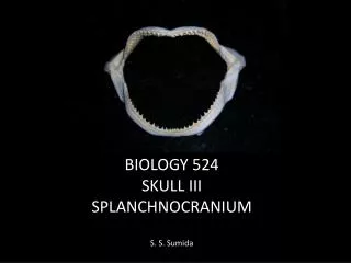
BIOLOGY 524 SKULL III SPLANCHNOCRANIUM S. S. Sumida. Gills

E N D
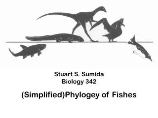
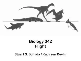
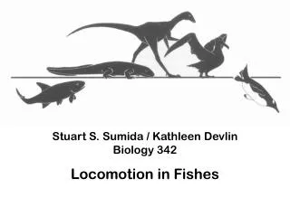
BIOLOGY 524 SKULL III SPLANCHNOCRANIUM S. S. Sumida
Gills The splanchnocranium is the skeleton of the gills. Gills of fishes are openings from the pharynx, through the body wall, to the outside of the body, allowing water to pass over the gills but leave the gut tube before heading to the remainder of the gut tube.
Embryology of the Splanchnocranium • All components of the vertebrate splanchnocraniumform endochondrally from neural crest cells. • Notably, whereas the entire splanchnocraniumis derived from neural crest forms endochondrally , it is not the only part of the skeleton derived from neural crest. • In general, fishes have a complete complement splanchnocranial elements. Through vertebrate evolution, elements of the first two arches, the mandibular arch (jaw) and the hyoid arch, have the greatest influence on skull construction.
SPLANCHNOCRANIUM OF JAWLESS VERTEBRATES In the most primitive jawless vertebrates, the head region is dominated by the gill apparatus. Some jawless vertebrates had as many as a dozen or even more gill openings. Note below the numerous gill openings of the agnathanHemiclaspis and how they dominate the mass of the head.
The heterostracanagnathanPterolepisshowing numerous gill openings. Individual components of the structure supporting the gill structures cannot be differentiated, so it is referred to as a BANCHIAL BASKET.
LIVING AGNATHANS Only two groups of living agnathans remain, the hagfishes and lampreys. They bear little resemblance to extinct agnathans, but they do retain the simple branchial basket as see above. Below: the branchial basket in the lamprey Petromyzon- a flexible catilagenous structure that can compress and expand under the influence of branchial musculature to help the animal ventilate its gills.
BAUPLAN PROGRESSION IN FISHES Note the change in organization of the gill structures.
Jaw Origin Hypothesis 1: There was an arch rostral to the one that became the jaws. It was subsequently lost, and is referred to as the premandibular arch. This is supported by the fact that part of the trigeminal nerve, the never of the jaw arch, or MANDIBULAR ARCH, has three major branches.. Most have two branches (one strictly sensory, the other sensory+motor). The trigeminal has an additional sensory branch reaching forward of the other two, and it is generally thought that this is the sensory branch of the old premandibular arch that was “captured” by the trigeminal. ORIGIN OF JAWS
Jaw Origin Hypothesis 2: Some researchers suggest there were two premandibular arches. But, as the arches are long gone,it is impossible to test. Jaw Origin Hypothesis 3: Some researchers suggest that not only was there a premandibular arch, but that there could have been another. This theory known as the COMPOSITE THEORY suggests that the resulting Mandibular arch as components of numerous adjacent arches incorporated. ORIGIN OF JAWS
In all of the hypotheses, at least one premandibular arch is suggested. The additional branch of the trigeminal nerve is cited as support for this hypothesis. Additional, many would suggest that the paired trabeculae of the rostral underside of the braincase are homologous to premandibular visceral arch elements. Immediately caudal to the mandibular arch, the next arch, known as the hyoid arch is also highly modified, in part to facilitate suspension of the jaws on the underside of the braincase. ORIGIN OF JAWS
OVERVIEW OF ORGANIZATION OF THE SPLANCHNOCRANIUM IN GNATHOSTOME FISHES: The vertebrate head is dominated by two great tubes: the dorsal hollow nerve tube, and the gut tube. The nerve tube’s influence on the skull derives from the protective function of the braincase and dermatocranial skull roof. The gut tube’s initial influence is due to the pharyngeal position of the gill sits, near to the head, and particularly the influence of the mandibular arch (jaws) and hyoid arch on skull structures.
OVERVIEW OF ORGANIZATION OF THE SPLANCHNOCRANIUM IN GNATHOSTOME FISHES: The vertebrate head is dominated by two great tubes: the dorsal hollow nerve tube, and the gut tube. The nerve tube’s influence on the skull derives from the protective function of the braincase and dermatocranial skull roof. The gut tube’s initial influence is due to the pharyngeal position of the gill sits, near to the head, and particularly the influence of the mandibular arch (jaws) and hyoid arch on skull structures.
SPLANCHNOCRANIUM IN CHONDRICHTHYES Cartilagenous fishes have no dermatocranium, and so their heads are dominated by the chondrocranium and the splanchnocranium. Elements of the splanchnocranium are essentially those of the illustration at the top of the page.
SPLANCHNOCRANIUM IN BONY FISHES In bony fishes, the mandibular arch, hyoid arch, and unmodified visceral arches are PRESENT. Separate ossifications point out that neither the upper jaw nor the lower jaw are single elements. Note particularly the jaw joint here is between the QUADRATE of the upper jaw and ARTICULAR of the lower jaw.
UPPER JAW ELEMENTS IN FISHES • Note the distal two most elements are the premaxilla and the maxilla. The neural crest components of these come to be covered by a sheath of intramembranous bone, and in more derived organisms they come to be incorporated into the lateral margin of the dorsal skull roof. (Those with * are later lost.) • Premaxilla • Maxilla • Palatine • Entopterygoid* • Ectopterygoid • Metapterygoid* • Quadrate • LOWER JAW ELEMENTS • Dentary • Articular • Note particularly the jaw joint her is between the quadrate and articular elements of the upper and lower jaws respectively.
JAW SUSPENSION IN FISHES The hyomandibula comes to play an important role in attaching the mandibular arch to the braincase. AUTOSTYLIC JAW SUSPENSION: Platoquadrate articulates with the underside of the skull. Probably the original condition for the earliest gnathostomes. AMPHISTYLIC JAW SUSPENSON: Upper jaw is suspended by both articulation with braincase and indirectly via hyomandibula. HYOSTYLIC JAW SUSPENSION: Jaws suspended by the hyoid arch only. (SECONDARY) AUTOSTYLIC JAW SUSPENSION: Platoquadrate (again) articulates with the underside of the skull.
THE LOWER JAW IN FISHES (AND OTHERS) The lower jaw in gnathostomes is a multi-element structure. The largest of the elements can be seen both laterally (externally, labially) and medially (internally, lingually). Directional Terminology along the tooth row cannot be simply cranially/rostrally versus caudally, as the jaw curves medially at its symphysis. So, proper terms are mesial (toward the symphysis) and distal. The story of the lower jaw is predominantly one of gradual loss or reduction in size of elements.
LATERALLY VISIBLE ELEMENTS OF LOWER JAW FROM MESIAL TO DISTAL: • Dentary (tooth bearing) • Splenial(s) (When two are present, sometimes referred to as splenial and postsplenial) • Angular • Surangular • MEDIALLY VISIBLE ELEMENTS OF LOWER JAW FROM MESIAL TO DISTAL: • Dentary (tooth bearing) • Coronoid(s) (tooth/fang bearing) • Prearticular • Articular • (Splenial(s) often visible at ventral margin)
SPLANCHNOCRANIUM IN BASAL TETRAPODS MANDIBULAR ARCH IN BASAL TETRAPODS: Ossifications of the upper jaw contribute to the skull. The epipterygoid is pieced out of the middle of the palatoquadrate cartilage. Mesially, the quadrate bone that has been incorporated into the posterolateral corner of the skull table provides the articulation with the lower jaw. This is the QUADRATE-ARTICULAR JAW JOINT. This jaw joint is retained through to modern extant amphibians.
The embryonic cartilage of the lower jaw is also known as MECKEL’S CARTILAGE. The lower jaw carries the full compliment of elements. The dentary is the primary, though not sole tooth-bearing bone. There are numerous elements in addition to the dentary, including the full compliment of splenials, coronoids, angular, surangular, parearticular, articular.
HYOID ARCH IN BASAL TETRAPODS – THE STAPES In basal tetrapods, the hyomandibula remains relatively large. Now known as the STAPES, it acts as a brace between the otic capsule of the braincase, and the inner surface of the dermal skull roof, inserting into a pit in the quadrate bone.
SPLANCHNOCRANIUM IN BASAL AMNIOTA • MANDIBULAR ARCH IN BASAL AMNIOTES: The condition of the jaw elements in basal amniotes is not significantly different from that of basal tetrapods. Chief differences include: • Reduction to single splenial. • Reduction to sincle coronoid. • Development of a RETROARTICULAR PROCESS, a caudally directed process of the elements of the distal end of the jaw to provide greater attachment for jaw opening muscles.
SPLANCHNOCRANIUM IN BASAL AMNIOTA HYOID ARCH IN BASAL AMNIOTES: The stapes remains as a brace between the otic capsule and the cheek region of the dermatocranium. Note the stapes is usually easy to identify due to the presence of the stapedial foramen.
JAW DIFFERENCES IN SELECTED REPTILIAN GROUPS: • In some herbivorous dinosaurs, the Cerapoda (which includes Ceratopsians such as Triceratops and inguanodonts), there is an additional bone forward of the dentary, the PREDENTARY. It is edentulous. • There is extreme reduction of jaw elements in snakes, and the lower jaws do not fuse at their symphysis to allow passage of prey. • Many reptiles have kinetic skulls to allow greater opening of the jaw, including a NASOFRONTAL HINGE in birds.
SPLANCHNOCRANIAL EVOLUTION IN SYNAPSIDA • The story of the transformation of the elements of the mandibular and hyoid components in the lineage leading toward mammals is perhaps one of the best known and most famous of evolutionary transformations taught in vertebrate biology. It is, briefly, the story of: • Consolidation of palatoquadrate elements into the side-wall of the braincase. • Reduction of post-dentary elements of the lower jaw. • Change from the quadrate-articular jaw joint to the DENTARY-SQUAMOSAL JAW JOINT. • Transformation of mandibular and hyoid arch elements into middle ear elements.
[Left – internal/lingual view; Right – external/labial view] • The dentary gradually becomes the dominant, then only bone of the lower jaw. • As it does so, it becomes (by default) the only tooth bearing bone. • The dentary develops a large, dorsally directed CORONOID PROCESS, which will take insertion of the TEMPORALIS muscle. • As this occurs, there is a progressive and proportional reduction in the size of the postdentary bones. • The splenial, coronoid, surangular, and angular are eventually lost. • The quadrate of the upper jaw, comes to be closely associated with the articular of the lower jaw – this is straight-forward, as they had already articulated as the quadrate-articular jaw joint of more primitive taxa.
The angular bone is reduced in size, detaches from the back of the jaw, attaches to the otic capsule of the skull, and becomes the circular rim of the EXTERNAL AUDITORY MEATUS of the temporal bone. • The articular is reduced in size. It remains lodged in the membrane that stretches across the articular rim of the external auditory meatus (TYMPANIC MEMBRANE), becoming the MALLEUS bone of the middle ear. • The quadrate is also reduced in size. It remains in articulation with the old quadrate (now the malleus), as the INCUS BONE. • The stapes is already in contact with the otic capsule. It comes to articulate with the incus.
Joint between incus and malleus = old quadrate-articular jaw joint. The angular contributes to the temporal bone, and the articular-quadrate-hyomandibular/stapes series become the MALLEUS-INCUS-STAPES linkage, or MIDDLE EAR OSSICLES.
COMPOSITE BONES OF THE MAMMALIAN SKULL • The bone most clearly explained by this discussion is the TEMPORAL BONE, which has the following components: • PETROUS portion – otic capsule of the chondrocranium • SQUAMOUS portion – squamosal of dermatocranium • ZYGOMATIC PROCESS – squamosal of dermatocranium • ANGULAR RIM of external auditory meatus – angular of mandibular arch of splanchnocranium • STYLOID PROCESS – hyoid arch
COMPOSITE BONES OF THE MAMMALIAN SKULL - II • OCCIPITAL BONE: • OCCIPITAL portion – Supraoccipital, exoccipitals, basioccipital of chondrocranium. • SQUAMOUS portion – postparietals of dermatocranium • Note the alisphenoid is in most mammals a separate ossification. • SPHENOID BONE: • GREATER WING (WHEN FUSED) – alisphenoid (= epipterygoid) of splanchnocranium • BASISPHENOID (including sellaturcica and dorsum sellae) – basisphenoid of chondrocranium • PTERYGOID WINGS – pterygoids of palatal dermatocranium • LESSER WINGS – Optic capsule of the chondrocranium