Download
1 / 58
580 likes | 784 Vues
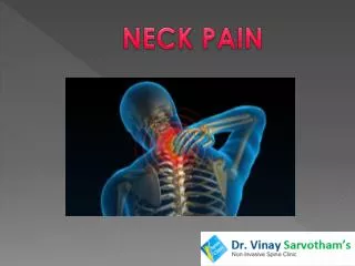
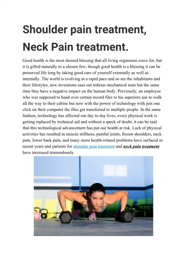
Neck pain is a common issue caused by various disorders, including cervical spondylosis, disc herniation, and trauma. This comprehensive overview discusses the anatomy of the cervical vertebrae, the mechanisms behind neck and upper extremity pain, and the role of conditions such as myelopathy and radiculopathy. We also cover treatment options, including medication, physical therapy, and surgical interventions when necessary. By understanding the causes of neck pain, patients can pursue effective management and regain function.

E N D
NECK PAIN Prof. Dr. Ece Aydoğ Yeditepe University Medical Faculty Department of PM&R
C1 or atlas There is nodiscbetween C1 and C2. 2.C2 oraxis 3.C3 4.C4 5.C5 6.C6 7.C7 8.Body 9.Vertebral foramen 10.Bifid spinousprocessorspine 11.Transverse process 12.Foramen transversariumortransverseforamen 13.Superior articularfacet Anatomy
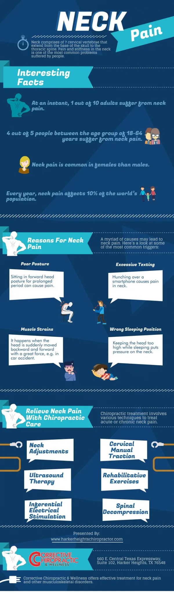
Disorders cause neck and upper extremity pain Cervical vertebra colon: cervical spondylosis (OA) cervical disc herniation spinal stenosis instability Wiplash injury Cervical cord diseases (Tumor, syringomyeli) Rheumatologic disorders: Ankylosing spondylitis, Rhematoid arthritis, Polymyalgia Rheumatica, Fibromyalgia, Myofascial pain syndrome Infectious: Osteomyelitis, dissit, epidural/intradural/subdural abces, retropharengeal abces Endocrin: Osteoporosis, osteomalasia, paget disease Trauma: sport injury, work conditions Thoracic outlet syndrome Neuropathies Artheritis (vertebral and cranial, Takayasu) Referred pain
Structures That Cause Neck Pain • Akromioclavicular joint • Heart and coronary disease • Apex of lung, Pancoast’s tumour • Diaphragm muscle (C3-C5 inn) • Gallbladder • Spinal cord tumour • Temporomandibular joint
Axial neck pain • Axial neck pain describes a pattern of pain that is localized to the occiput and neck region • Degenerative arthritis within the upper cervical spine can manifest as suboccipital headache and localized pain. This is termed cervicogenic headacheand is thought to result from irritation of the greater occipital nerve
Cervical strain and sprain • Strain: • injury of contractile tissues by stretching (muscle, lig.) • Pain is localized on neck • Decreased lordosis, paravertebral spasm • No neurologic sign • Sprain: • Tissue rupture and bleeding by stretching (capsule, lig., bursa, vessels, cartilage, dura)
Cervical spondylosis • Degeneration of IVD, facet and luschka • Age, microtrauma, ergonomy, genetic • Syndromes due to spondylosis • Radiculopathy • Vertebrobasilar insufficiency • Cervical myelopathy
Radiculopathy • Radicular pain • Paresthesias • Superficial sensory deficits • Variation of DTR • Muscle strength loss
If these deficits are minor and tolerable, it is reasonable to treat with conservative care with close follow-up to ensure that the deficit is not progressive • Disabling deficits should be treated operatively because prolonged nerve compression can result in irreversible changes • In patients without a neurologic deficit, it is reasonable to expect a good outcome with conservative care
Myelopathy • Myelopathy is the clinical presentation of long tract signs resulting from compression of the spinal cord • Myelopathy: • tumor or infection • instability owing to systemic arthritides or connective tissue disorders • degenerative changes within the cervical spine • Factors that contribute to the development of myelopathy : • congenitally narrow spinal canal, dynamic cord compression, dynamic thickening of the spinal cord, and vascular changes
complaints of hand clumsiness or difficulty with balance • worsening handwriting or difficulty buttoning buttons • nausea and emesis caused by equilibrium dysfunction • paresthesias and dysesthesias may be present, often involving bilateral upper extremities and not following a dermatomal distribution • wasting of hand intrinsics and bowel and bladder dysfunction
Definitive indications for surgery: • presence of myelopathy for 6 months or longer, • progression of signs or symptoms, • difficulty walking, or change in bowel or bladder function.
Vertebrobasiller Insufficiency • Blood supply of inner ear, vestibular and cochlear nucleii of medulla oblangata • Vertigo, headache, nausea • Coordination, memory deficit • Tinnitus, hearing loss, diplopia • Nistagmus, disphagia • Common property of those symptoms is that they are related with neck movement and local/radicular symptoms
Cervical disc herniation • With age, the nucleus pulposus becomes vulnerable • With degenerative changes, • the disc space narrows, spinal column shortens • The intervertebral foramina become narrowed, movements become restricted, unusual mechanical strains on the sinovial joints result • The formation of osteophytes leads to encroachment on the spinal canal and intervertebral foramina • Changes in the caliber of the vertebral arteries can result because of the degenerative changes • Facet joints (sinovial) can be affected by various arthritic diseases
Treatment • Immobilization: cervical collar • Medication: NSAID, analgesics, myorelexan, corticosteroids • Physical therapy: superficial and deep heat, massage, electrotherapy, traction • Theurapatic exercises: Isometric, ROM
Thoracic outlet syndrome • Thoracic outlet syndrome is a condition whereby symptoms are produced from compression of nerves or blood vessels, or both, because of an inadequate passageway through an area (thoracic outlet) between the base of the neck and the armpit • muscle enlargement (such as from weight lifting), injuries • an extra rib from the neck at birth (cervical rib) • weight gain • tumors at the top of the lung (rare) • often no specific cause is found
Anatomic regions causing compression: 1. Interscalenetriangle 2. costaclavicular fossa
Wilbourne classification Vascular %10 Neurogenic %90 Real neurogenic: • C8-T1 pain and paresthesia • Generalised painarm, anterior and posterior chest wall • Atrophy and muscle weakness at hand Suspicious neurogenic: • Same symptoms but no objective signs
Symptoms • neck, shoulder, and arm pain • numbness, or impaired circulation to the extremities (causing discoloration) • often symptoms are reproduced when the arm is positioned above the shoulder or extended • pains can extend to the fingers and hands, causing weakness
Provocative tests Adsontest Costoclavicular compression test Roos test
Cervical graphy: • Cervical costa • PA lung graphy: Pancoast tm • ENMG: although these may not be positive in all patients • Angiogram x-ray tests:demonstrate the pinched area of the blood vessel involved.
Treatment Conservativetreatment • Postural exercises • Shoulder girdle strengthening exercises and scalene muscles streching exercises • Myorelaxan • NSAİİ • Superficial and deep heat, iontophoresis,TENS Surgery
avoid prolonged positions with their arms held out or overhead • avoid sleeping with the arm extended up behind the head • have rest periods at work to minimize fatigue • weight reduction • avoid sleeping on their stomach with their arms above the head • not repetitively lift heavy objects
Anatomy And Function • The shoulder joint is the most mobile joint of the body • Joint stability: • Labrum, capsule and the glenohumeral ligaments (static stability) • Rotator cuff (dynamic stability) • The shoulder consists of three joints; • Acromioclavicular (AC) • Sternoclavicular • Glenohumeral • (Two gliding planes: scapulothoracic and subacromial surfaces)
Intransic causes: Impingement Calcific tendinits Frosen shoulder Biceps tendinitis Glenohumeral instability Degeneration Arthritis Avascular necrosis Fracture tumor Extransic causes: Cervical radiculopathy Bracial neurit, bracial plexus injury Toracic outlet syndrome Cardiac referred pain Abdomial referred pain Tumor Fracture Fibromiyalgia Myofacial pain syndrome Shoulder Pain Causes
Bicipital Tendinitis And Rupture • The biceps tendon aids in flexion of the forearm, supination of the pronated forearm if the elbow is flexed, and forward elevation of the shoulder. • Anterior shoulder pain • Yergason's supination sign • Treatment generally is conservative and consists of rest, analgesics, NSAIDs, and local injection of glucocorticoids. • Patients with refractory bicipital tendinitis and recurrent symptoms of subluxation are treated by arthroscopic interventions
Impingement • Impingement : encroachment of the acromion, coracoacromial ligament, coracoid process, or AC joint on the rotator cuff as it passes beneath them during glenohumeral motion • The mechanical impingement : shape and slope of the acromion, proliferative spur formation of the acromion or degenerative changes of the AC joint • Stage I lesion: • Edema and hemorrhage • Conservative treatment • Stage II lesions: • Fibrosis and thickening of the tendon after repeated episodes of mechanical impingement over time • Treated conservatively, but attacks may recur • If symptoms persist despite adequate conservative management for more than 6 to 12 months, surgical intervention is warranted. • Stage III lesions: • Rotator cuff tears, biceps tendon rupture, and bone changes • Pain, weakness, or supraspinatus atrophy • Surgical treatment
Pain: located over the anterior and lateral aspects of the shoulder and may radiate into the lateral deltoid • The pain may worsen with sleeping on the affected extremity and is exacerbated by overhead activity • Tenderness on palpation: greater tuberosity bicipital groove, AC joint • The impingement sign: Neer test, Yergason test • Radiographs • Arthrography, MRI, and ultrasound
Rotator Cuff Tear • Inflammatory diseases • Metabolic conditions: Renal osteodystrophy • Glucocorticoids • Trauma: falling on an outstretched arm or lifting a heavy object • Pain and weakness of abduction and external rotation • Atrophy of the supraspinatus and infraspinatus muscles • Drop arm test
Conservative treatment • The mainstay of conservative therapy is exercise • Rehabilitation stresses pain relief with exercises aimed at restoring shoulder motion and strengthening the remaining cuff muscles, deltoid, and scapular stabilizers • Steroid and local anesthetic injections • If the patient fails to improve after 3 months of conservative treatment, or does not continue to improve after three sequential injections, surgical options should be discussed
Calcific Tendinitis • The cause: degeneration of the tendon, which leads to calcification through a dystrophic process • A common clinicopathologic correlation is three distinct phases of the disease process: • Precalcific or formative phase: relatively painless • Calcific phase: which tends to be quiescent and may last months to years • Resorptive orpostcalcific phase: painfulphase • Impingement-type pain • The acute inflammation can be treated with local glucocorticoid injection, NSAIDs, or both
Adhesive Capsulitis • Adhesive capsulitis, or frozen shoulder syndrome (FSS), is a condition characterized by limitation of motion of the shoulder joint with pain at the extremes of motion • Diabetes mellitus • Parkinsonism • Thyroid disorders • Cardiovascular diseases
Capsular contracture is thought to result from adhesion of the capsular surfaces or fibroblastic proliferation in response to cytokine production • More common in women in their 40s and 50s • Diffuse, dull aching around the shoulder • Muscle weakness • Loss of motion
Stage 1:Painful or freezing phase • Stage 2: Adhesive or stiffening phase and generally lasts 4 to 12 months • Stage 3:Resolution or thawing phase and may last 5 to 26 months
In patients with a history of minimal or no trauma and FSS, a metabolic cause should be excluded • Complete blood cell count • Erythrocyte sedimentation rate • Serum chemistry • Thyroid function tests
Osteonecrosis • The most common cause of osteonecrosis of the shoulder is avascularity resulting from a fracture through the anatomic neck of the humerus • Steroid therapy in conjunction with organ transplantation,systemic lupus erythematosus, or asthma • Other conditions: hemoglobinopathies, pancreatitis, and hyperbarism.
Milwaukee shoulder • Deposition of calcium pyrophosphate dihydrate crystals, direct trauma, chronic joint overuse, chronic renal failure, and denervation • Hemiarthroplasty or a reverse total shoulder arthroplasty may be indicated
Labral Tears • Bankart lesion • SLAP lesion
Carpal Tunnel Syndrome Carpal Tunnel Syndrome is a nerve disorder in the hand that causes pain and loss of feeling, especially in the thumb and first 3 fingers.
Tingling or numbness in part of the hand. Sharp pains that shoot from the wrist up the arm, especially at night. Burning sensations in the fingers. Morning stiffness or cramping of hands. Thumb weakness. Frequent dropping of objects. Inability to make a fist. Shiny, dry skin on the hand. Signs and Symptoms