Download
1 / 40
421 likes | 529 Vues
Explore the components, circulation, and significance of fetal membranes and amniotic fluid during pregnancy. Learn about the functions and importance of the yolk sac and allantois in fetal development.

E N D
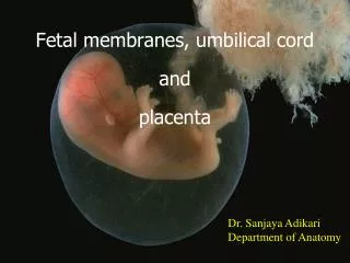
Fetal Membranes Dr. Mujahid Khan
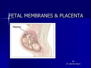
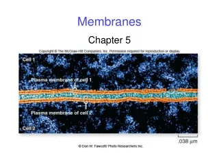
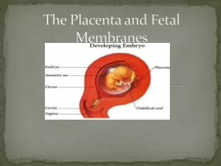
The fetal part of the placenta and fetal membranes separate the fetus from the endometrium of the uterus • An interchange of substances such as nutrients and oxygen occurs between the maternal and fetal blood streams through the placenta
What constitute a Fetal Membrane • Decidua • Chorion • Amnion • Yolk sac • Allantois
Amnion • Thin but tough • Forms a fluid filled membranous amniotic sac that surrounds the embryo and fetus • Is attached to the margins of the embryonic disc • Its junction with embryo located on the ventral surface after the folding
Amniotic Fluid • Plays a major role in fetal growth and development • Most of it is derived from maternal tissue and by diffusion across the amniochorionic membrane from the decidua parietalis • Later there is a diffusion of fluid through the chorionic plate from blood in the intervillous space of the placenta
Amniotic Fluid • Amniotic fluid is similar to fetal tissue fluid • Before keratinization of the skin the pathway for passage of water and solutes in tissue fluid from the fetus to the amniotic cavity is through the skin • Fluid is also secreted by the fetal respiratory tract and enters the amniotic cavity
Amniotic Fluid • Daily contribution of fluid from respiratory tract is 300-400 ml • Fetus contributes to the amniotic fluid by excreting urine into the amniotic cavity • Half a liter of urine is added daily during the late pregnancy • Amniotic fluid volume is 30 ml at 10 weeks, 350 ml at 20 weeks, 700-1000 ml at 37 weeks
Circulation of Amniotic Fluid • Water content of amniotic fluid changes every 3 hours • It is been swallowed by the fetus and absorbed by respiratory & digestive tracts • Fetus swallows up to 400 ml of fluid per day during the end days of pregnancy
Circulation of Amniotic Fluid • Fluid passes into the fetal blood stream and the waste products in it cross the placental membrane and enter the maternal blood in the intervillous space • Excess water in the fetal blood is excreted by the fetal kidneys and returned to the amniotic sac as a urine
Disorders of Amniotic Fluid Volume • Oligohydromnios • Renal agenesis • Obstructive uropathy • Polyhydromnios • Esophageal atresia
Exchange of Amniotic Fluid • Large amount of amniotic fluid move in both directions between the fetal and maternal circulations mainly through the placental membrane • Most fluid passes into GIT but some passes into lungs • Fluid is absorbed in either case and enters the fetal circulation • It then passes into the maternal circulation through the placental membrane
Composition of Amniotic Fluid • 99 % is water • Desquamated fetal epithelial cells • Organic & inorganic salts • Protein, carbohydrates, fats, enzymes, hormones • Meconium & urine in the late stage • Amniocentesis can be performed to check the concentration of different compounds for diagnostic purpose
Composition of Amniotic Fluid • High levels of alpha-phetoprotein (AFP) in amniotic fluid usually indicate the presence of a severe neural tube defect (meroanencephaly) • Low levels of AFP may indicate chromosomal aberrations such as trisomy 21
Significance of Amniotic Fluid • Permits symmetrical external growth of the embryo and fetus • Acts as a barrier to infection • Permits normal fetal lung development • Prevents adherence of amnion to fetus • Cushions & protects the embryo and fetus • Helps maintain the body temperature • Enables the fetus to move freely
Yolk Sac • It is large at 32 days • Shrinks to 5mm pear shaped remnant by 10th week & connected to the midgut by a narrow yolk stalk • Becomes very small at 20 weeks • Usually not visible thereafter
Significance of Yolk Sac • Has a role in transfer of nutrients during the 2nd and 3rd weeks • Blood development first occurs here • Incorporate into the endoderm of embryo as a primordial gut • Primordial germ cells appear in the endodermal lining of the wall of the yolk sac in the 3rd week
Fate of Yolk Sac • At 10 weeks lies in the chorionic cavity between chorionic and amniotic sac • Atrophies as pregnancy advances • Sometimes it persists throughout the pregnancy but of no significance • In about 2% of adults the proximal intra-abdominal part of yolk stalk persists as an ileal diverticulum or Meckel diverticulum
Allantois • In the 3rd week it appears as a sausagelike diverticulum from the caudal wall of yolk sac that extends into the connecting stalk • During the 2nd month, the extraembryonic part of the allantois degenerates
Functions of Allantois • Blood formation occurs in the wall during the 3rd to 5th week • Its blood vessels persist as the umbilical vein and arteries • Fluid from the amniotic cavity diffuses into the umbilical vein and enters the fetal circulation for transfer to maternal blood through placental membrane • Becomes Urachus and after birth is transformed into median umbilical ligament extends from the apex of the bladder to the umbilicus
Umbilical Cord • Is attached to the placenta usually near the center of the fetal surface of this organ • May attach to any other point • Is usually 1-2 cm in diameter and 30-90 cm in length • Long cord may cause prolapse or compression of the cord which may lead to fetal hypoxia • Short cord may cause premature separation of the placenta from the wall of the uterus during delivery
Umbilical Cord • Has two arteries and one vein surrounded by Wharton jelly • Umbilical vessels are longer than the cord, so twisting and bending of the vessels are common • They frequently form loops, producing false knots, that are of no significance • In about 1% of pregnancies, true knots form in the cord and cause fetal death
Chorion • Primary chorionic villi appear by the end of the 2nd week • Growth of these extensions are caused by underlying extraembryonic somatic mesoderm • The cellular projections form primary chorionic villi
Chorion • The extraembryonic somatic mesoderm and the two layers of trophoblast form the chorion • Chorion forms the wall of chorionic sac • Embryo and its amniotic and yolk sacs are suspended into it by connecting stalk • The extraembryonic coelom is now called the chorionic cavity
Chorion • The amniotic sac with embryonic epiblast form its floor • The yolk sac with embryonic hypoblast form its roof • Are analogous to two balloons pressed together, suspended by a connecting stalk from the inside of a larger balloon (chorionic sac)
Chorion • Transvaginal ultrasound is used to measure the chorionic sac diameter • This measurement is valuable for evaluating the early embryonic development and pregnancy outcome
Chorion • Chorionic villi cover the entire chorionic sac until the beginning of 8th week • As this sac grows, the villi associated with decidua capsularis are compressed, reducing the blood supply to them • These villi soon degenerates producing an avascular bare area smooth chorion (chorion laeve)
Chorion • As the villi disappear, those associated with the decidua basalis rapidly increase in number • Branch profusely and enlarge • This bushy part of the chorionic sac is villous chorion
Decidua • The gravid endometrium is known as decidua • It is the functional layer of endometrium in a pregnant woman • This part of the endometrium separates from the rest of the uterus after parturition
Regions of Decidua 3 regions of decidua are: • Decidua basalis: lies deep to the conceptus that forms maternal part of the placenta • Decidua capsularis: superficial part that overlies the conceptus • Decidua parietalis: is all the remaining parts of the decidua
Decidua • In response to increasing progesterone levels in the maternal blood the connective tissue cells of the decidua enlarge to form decidual cells • These cells enlarge as glycogen and lipid accumulate in their cytoplasm
Decidua • The cellular and vascular changes occurring in the endometrium as the blastocyst implants constitute the decidual reaction • Many decidual cells degenerate near the chorionic sac in the region of the syncytiotrophoblast • Together with maternal blood the uterine secretions provide a rich source of nutrition for the embryo
Decidua • The full significance of decidual cells is not understood • They may protect the maternal tissue against uncontrolled invasion by the syncytiotrophoblast • They may be involved in hormonal production • Clearly recognizable during ultrasonography to diagnose early pregnancy