Download
1 / 29
310 likes | 367 Vues
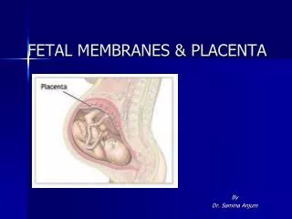
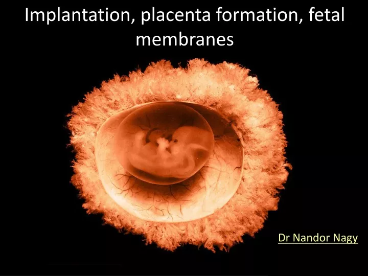
Implantation, placenta formation, fetal membranes. Dr Nandor Nagy. By the 5th to 6th day of development, the embryo is a hollow ball of about 100 cells called a blastocyst. At this point, it enters the uterine cavity and begins to implant into the endometrial lining of the uterine wall.

E N D
Implantation, placenta formation, fetal membranes Dr Nandor Nagy
By the 5th to 6th day of development, the embryo is a hollowball of about 100 cells called a blastocyst. At this point, it enters the uterine cavity and begins to implant into the endometrial lining of the uterine wall.
CLEAVAGE early compact morula 8 cell-stage morula pronuclei late morula blastocyst hatching
DECIDUAL REACTION Very soon after arriving in the uterus, the blastocystbecomes tightly adherent to the uterine lining. The adjacent cells of the endometrial stroma respond to its presence and to the progesteronesecreted by the corpus luteum by differentiating into metabolically active, secretory cells called decidualcells.
HUMAN CHORIONIC GONADOTROPIN (hCG) the trophoblast produce the hormone human chorionic gonadotropin (hCG), which supports the corpus luteum and thus maintains the supply of progesterone (maternal recognition of pregnancy).
Between days 6 and 9, the embryo becomes fully implanted in the endometrium
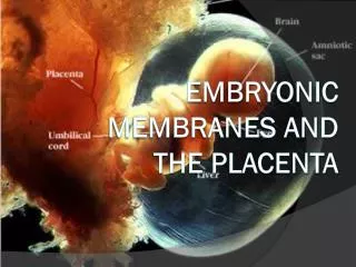
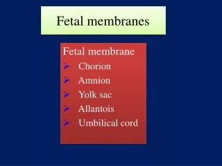
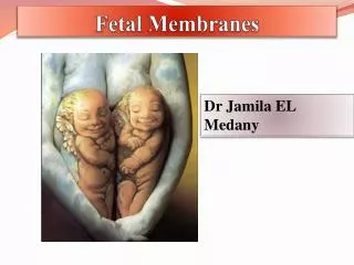
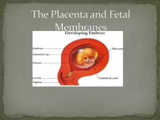
4 extraembryonic membranes: Amnion, Yolk Sac, Chorion, Allantois….
Amnion At the beginning of the 2nd week, the embryoblast splits into two layers, the epiblast and the hypoblast, or primitive endoderm.
Although the amniotic cavity is at first smaller than the blastocystcavity, it expands steadily. By the 8th week, the amnionencloses the entire embryo.
Chorion extraembryonic coelom, or chorionic cavity—developsas the extraembryonic mesoderm splits into two layers.
Yolk sac By day 12, the primary yolk sac is displaced(and eventually degenerates) by the second wave ofmigrating hypoblast cells, which forms the secondary yolk sac.
End of the 2nd week The syncytiotrophoblast, cytotrophoblast, and associated extraembryonic mesoderm, together with the uterus, initiate formation of the placenta. During this process, the fetal tissues establish outgrowths,the chorionic villi, which extend into maternal blood sinusoids.
By day 13, the embryonic disc with itsdorsal amnion and ventral yolk sac is suspended inthe chorionic cavity solely by a thick stalk ofextraembryonic mesoderm called the connecting stalk.
yolk sac functions: -Extraembryonic mesoderm forming the outer layerof the yolk sac is a major site of hematopoiesis. -Primordial germ cells canfirst be identified in humans in the wall of the yolksac. After the 4th week, the yolk sac is rapidlyovergrown by the developing embryonic disc. The yolk sac normally disappears before birth,but on rare occasions it persists in the form of a digestivetract anomaly called Meckel’s diverticulum.
The portion of the primitive hindgut tube lying justdeep to the cloacal membrane forms an expansioncalled the cloaca. A slim diverticulum of the cloacacalled the allantois extends into the connecting stalk. It helps the embryo exchange gases and handle liquid waste. Allantois
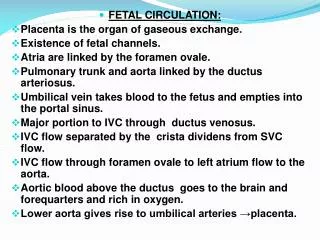
Placenta formation During the 1st week of development, the embryoobtains nutrients and eliminates wastes by simple diffusion.Rapid growth of the embryo makes a more efficientmethod of exchange imperative. This need is filled bythe uteroplacental circulation—the system by whichmaternal and fetal blood flowing through the placentacome into close proximity and exchange gasesand metabolites by diffusion. This system begins toform on day 9 as vacuoles called trophoblastic lacunaeopen within the syncytiotrophoblast.
Maternal vessels near the syncytiotrophoblast expand to form maternal sinusoids that rapidly anastomose with the trophoblastic lacunae. Between days 11 and 13, asthese anastomoses continue to develop, the cytotrophoblast proliferates locally to form extensions that growinto the overlying syncytiotrophoblast.
By the end of the 3rd week, this villous mesoderm hasgiven rise to blood vessels that connect with the vesselsforming in the embryo proper, thus establishing a working uteroplacental circulation. Villi containing differentiated blood vessels are calledtertiary chorionic stem villi.
Placenta fetalblood - maternalbloodbarrier The gases, nutrients, andwastes that diffuse between the maternal and fetalblood must cross four tissue layers: the endothelium of the villus capillaries the loose connective tissue in the core of the villus (extraembryonic mesoderm) a layer of cytotrophoblast a layer of syncytiotrophoblast Mesenchyme Trophoblast fetal vessels Precisely: Syntitiotrophoblast (Cytotrophoblast) Membrana basalis Mesenchyme Membrana basalis Endothel Intervillous space maternal blood
Umbilicalcord Umbilical arteries Amnion Wharton- jelly Left umbilical vein
hydatidiform mole, which is characterized by the overdevelopment of trophoblastic tissues and the extreme underdevelopment of the embryo.This condition can result from the fertilization of an egg by two spermatozoa and the consequent failure of the maternal genome of the egg to participate in development or from the duplication of a sperm pronucleus in an "empty" egg.