Download
1 / 77
810 likes | 1.2k Vues
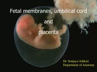
Fetal membrane and placenta. Decidua: After the implantation of the embryo, the uterine endometrium is called the decidua. The stramal cells enlrge,become vacuolated and lipids.This change in the stromal cells is called the decidua reaction.

E N D
Decidua: • After the implantation of the embryo, the uterine endometrium is called the decidua. The stramal cells enlrge,become vacuolated and lipids.This change in the stromal cells is called the decidua reaction.
The decidua is divided into three parts according to the association with the embryo: Decidua basalis: deep to the embryo Decidua capsulris: over the embryo Decidua paritalis: the left part of decidua
Decidua capsulris Decidua basalis Decidua paritalis
Fetal membrane: • Chorion • Amnion • Yolk sac • allantois • Unbiliad cord
Lacuna syncytiotrophoblast cytotrophoblast Extraembryonic mesoderm
Chorion: Formation of chorion:The cytotrophoblast differentiates internally into a layer of primary mesoderm. Trophoblast and primary mesoderm together form the chorion.They give off numerous process called villi or chorionic villi.These villi are surrounded by maternal blood .
The chorionic villi are first formed all over the trophoblast and grow into the surrounding decidua.those related to the decidua capsularis are transitory. After some time they degenerate.This part of the chorion becomes smooth and is called the chorion laevae.
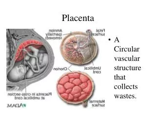
In contrast,decidua undergo considerable development. Along with the tissues of the decidua basalis these villi form a disc-shaped mass which is called the placenta. The part of the chorion that helps form the placenta is called the chorion frondosum.
Amnion Connecting stilk Germ disc Yolk sac chorion
Amnion Connecting stilk Yolk sac Extraembryonic cavity
Extraembryonic cavity Amniotic cavity Umbilical cord Yolk sac
Amniotic cavity Umbilical cord amnion Chorion laevae
All elements (syncytium, cytotrophoblast and mesoderm) take part in forming chorionic villi.Three stages in formation of chorionic villi are seen: A primary villus:cytotrophoblast and is covered by the cells of syncytiotrophoblast.
A secondary villus:primary mesoderm and is covered successively by cyto-and syncytiotrophoblasts. • A tertiary villus contains in the center the foetal blood vessels which are surrounded successively from within outwards by primary mesoderm, cyto and syncytiotrophoblasts.
capillary Intervilli space Cell shell decidua Syncytiotrophoblast Connective tissue capillary cytotiotrophoblast
From each tertiary stem villus numerous branching villi project into the intervillous space. the intervillous space is converted into a sponge-like network of villous type of labyrinthine structure and is filled with maternal blood.
THE AMNIOTIC CAVITY • A fluid-filled amniotic cavity appears in the second week of development between the germ disc and the trophoblast. • The roof of the cavity: a layer of flattened cells, the amnioblast, which lines the inner aspect of the cytotrophoblast. • The floor of the cavity:the tall columnar cells of the epiblast of the germ disc.
Epiblast Aminiotic cavity Hypioblast
With the extension of the extra-embryonic coelom the outer surfaces of the amniotic cavity and the yolk sac are covered with a layer of primary mesoderm, which is continuous with the primary mesoderm of the chorion at the caudal end of the germ disc through the connecting stalk.
The formation of the embryonic folds allows the amniotic cavity to surround the outer surface of the cylindrical embryo. As a result the amnio-ectodermal junction converges towards the ventral surface of the embryo to form the umbilical cord
The amniotic cavity gradually increases in size at the expense of the extra-embryonic coelom, and eventually the amnion and chorion leave are fused. Finally the extra-embryonic coelom is completely obliterated, except a small part which is contained in the proximal part of the umbilical cord up to the 10th week of intra-uterine life.
Amnion Connecting stilk Germ disc Yolk sac chorion
Amnion Connecting stalk Yolk sac Extraembryonic cavity
Extraembryonic cavity Amniotic cavity Umbilical cord Yolk sac
Amniotic cavity Umbilical cord amnion Chorion laevae
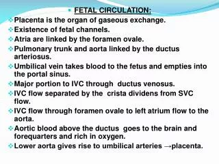
The Amniotic Fluid • The amniotic fluid, also called liquor amnii, is clear and watery, containing about 2% solids which include inorganic salts, urea, proteins and a trace of sugar. The source of the fluid still remains unsettled-it may be foetal from the amniotic cells, maternal or both.
Functions of the liquor amnii- • 1.It acts as a protective cushion for the embryo against shock, blows or pressure. The embryo is suspended by the umbilical cord and literally swims in the fluid. • 2.The fluid maintains a uniform pressure for the proper growth and differentiation of the delicate tissues of the embryo.
3.It allows foetal movements and maintains a constant environmental temperature. • 4.During parturition, the amniotic sac forms a hydrostatic wedge and helps to dilate the cervical canal.
Abnormalities of the liquor amnii- • 1.Hydramnios This is a condition when the volume of the amniotic fluid exceeds two litres. • 2.Oligamnios- In this condition the fluid is scanty in amount.
Yolk sac Primary Yolk sac secondary yolk sac
Epiblast Aminiotic cavity Hypioblast
allantois Primitive female sex cell Yolk sac
.Vitello-intestinal duct –It communicates the mid gut with extra-embryonic part of the yolk sac (umbilical vesicle). In the later part of foetal life the duct disappears. On rare occasions, the proximal part of the duct persists as the Meckel’s diverticulum which is attached to the antimesenteric border of the ileum.