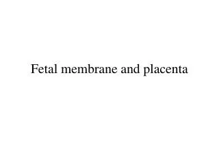
EXTRAEMBRYONIC MEMBRANE AND PLACENTA
EXTRAEMBRYONIC MEMBRANE AND PLACENTA. PREPARED BY: WILSON R. JACINTO. Emb ryonic stem cells from inner cell mass to produce restricted stem cell to produce neurons and blood. Universal requirement - embryo to develop in moist, protective environment Adaptations developed by vertebrates

EXTRAEMBRYONIC MEMBRANE AND PLACENTA
E N D
Presentation Transcript
EXTRAEMBRYONIC MEMBRANE AND PLACENTA PREPARED BY: WILSON R. JACINTO
Emb ryonic stem cells from inner cell mass to produce restricted stem cell to produce neurons and blood
Universal requirement - embryo to develop in moist, protective environment • Adaptations developed by vertebrates • Laying and fertilizing eggs in water (oviparous) • Incubation of embryo internally and give live birth (viviparous) • Eggs incubated internally and hatch just in time for release from the mother’s body (ovoviviparous) • For nutrition and exchange of gas
elaboration of a protective shell and a series of cellular membranes surrounding the embryonic body - A significant evolutionary step that enable reptiles to lay eggs capable of developing on land. • Vital functions of the membranes • Keep embryo in fluid • Gas exchange • Removal or storage of waste materials • Nutrition
Four Sets of Embryonic Membranes in Land Vertebrates • Membranous folds formed by extension of ectoderm and endoderm underlain with lateral plate mesoderm • Somatopleure – forms amnion and chorion • Splanchnopleure – allantois and yolk sac
Amnion – thin ectodermally-derived membrane enclosing embryo in fluid-filled sac • For secretion and absorption of amniotic fluid • Yolk sac – endodermal, • involved with nutrition of the embryo in large–yolked forms.
Allantois – endodermal, originate from ventral surface of early hindgut; • Act as reservoir for storing or removing urinary wastes and mediate gas exchange between embryo and surrounding • Chorion – outermost membrane; • In species that lay eggs, the principal function is the respiratory exchange of gases. • In mammals, nutrition, excretion, filtration and synthesis (hormone)
Extraembryonic Membrane of the Chick – Yolk Sac • splanchnopleure grows over yolk surface; yolk as temp. floor; then completely surround the yolk • Subcephalic pocket - forms floor of foregut • Subcaudal pocket – hindgut • Midgut • Yolk duct – opening of the midgut • Yolk stalk – wall of the duct • Vitelline arc – yolk circulatory arc • Endoderm of yolk sac – absorptive and synthetic • Completely resorpted 6 days after hatching
Amnion and chorion • Derived from extraembryonic somatopleure • First indication appears about 30 hours of incubation • Somatopleure is thrown into a head fold and tail fold and lateral folds • Continued growth of the amnion results in the meeting above the embryo, results to scar –like thickening called amniotic raphe
The outer layer of somatopleure becomes the chorion • Chorioamniotic cavity • Rapid peripheral growth of the somatopleure carries the chorion about the yolk sac, which it eventually envelopes • Thus, chorion eventually encompasses the embryo itself and all other membranes. • Important function – transport of Ca++ from shell to embryo
Allantois • Arises from the endoderm of the ventral wall of the hindgut • Appears late in the third day of incubation • Pushes out to the extraembryonic coelom (chorioamniotic cavity) • Grows rapidly and finally encompasses the embryo and yolk sac • The mesodermal layer of allantois fuse with mesodermal layer of chorion.
The double-layer of mesoderm develops into vascular network connected to the embryo by allantoic veins and arteries • Primary function is oxygenating blood and relieve carbon dioxide • Fuse the chorion – chorioallantoic membrane • Also, reservoir for secretions coming from developing excretory organs • Urea in early stages; uric acid later stages
Formation of Extra-embryonic membranes and Placenta in mammals • Mammals form and utilize the same extraembryonic structures as does the chick with some modifications • Trace the development of the four extraembryonic membranes in mammals
Amnion • Primordial amniotic cavity forms by means of cavitation within epiblastic components of inner cell mass • Cytotrophoblast bounds the primordial amniotic cavity, then later by the spreading of the epiblast • Epiblast becomes surrounded by the spreading extraembryonic mesodermal cells derived from primitive streak
Yolk Sac • Hypoblast , which segregated from the inner cell mass spread to the inner surface of trophoblast – constitute the primary yolk sac • Extraembryonic mesoderm (splanchnic mesoderm) arise from some hypoblast and caudal margin of primitive streak. • Secondary yolk sac is formed as the primitive yolk sac collapses.
Chorion • Trophoblast cells expands and forms masses suggestive of the villi – primary villi (2nd week) • As the chorionic vesicle receives ingrowth of allantoic vessels and mesoderm – secondary villi • Villi with a vascular connective tissue core - tertiary villi (3rd week) • Tertiary villi is retained throughout pregnancy
Tertiary chorionic villi contain embryonic blood vessels that develop to loose connective tissue core. The vessels connect with vessels in the chorion and connecting stalk and begin to circulate embryonic blood about the third week of development.
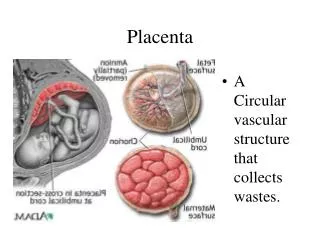
Placenta Uterine stromal cells around blastocyst - enlarge, form glycogen and lipid droplets -- decidual reaction, decidual cells The endometrium (lining of the uterus) of the mother consisting of three regions named by location.
Deciduacapsularis become attenuated and atrophy at the end of trimester – becomes smooth • chorionlaeve • Chorionfrondosum – with highly developed villi • Interlocked chorionfrondosum of fetus and anddeciduabasalis = placenta
In mammalian embryo, food and oxygen are obtained through the placenta – lack of functional lungs and intestines • Allantoic (umbilical) mesoderm and blood vessels spread out along chorion – forms placenta • Connected to the umbilical vein (carries oxygenated and food-laden blood) of the embryo • Umbilical arteries carry wastes to the placenta • Types: epitheliochorial, syndesmochorial, endotheliochorial and hemochorial
Fetal hemoglobin has higher affinity to oxygen than adult hemoglobin • Myoglobin of fetal muscle has even greater affinity – thus stores oxygen
Oxygen and nutrients in the maternal blood in the intervillous spaces diffuse through the walls of the villi and enter the fetal capillaries. • Carbon dioxide and waste products diffuse from blood in the fetal capillaries through the walls of the villi to the maternal blood in the intervillous spaces.
Amniotic fluid • Amniotic fluid main functions: • protects the fetus physically, • room for fetal movements, and • regulate fetal body temperature. • produced by dialysis of maternal and fetal blood through blood vessels in the placenta. • Later, production of fetal urine contributes to the volume of amniotic fluid and fetal swallowing reduces it. The water content of amniotic fluid turns over every three hours.
Umbilical cord • A composite structure formed by contributions from: • Fetal connecting (body) stalk • Yolk sac • Amnion • contains the right and leftumbilicalarteries, the left umbilical vein, and mucous connective tissue. Presence of only one umbilical artery may suggest the presence of cardiovascular anomalies.
Fetal Circulation • Fetal circulation involves three circulatory shunts: the ductus venosus, which allows blood from the placenta to bypass the liver, and the ductus arteriosus and foramen ovale, which together allow blood to bypass the developing lungs. Refer to the section on changes at birth for more information on the fates of these structures.
Ductus arteriosus diverts blood from pulmonary artery to aorta Foramen ovale – hole in septum between atria. This brings blood to ventricle - may remain open
Clinical Correlations • Multiple Pregnancy • Dizygotic twins - derived from two zygotes that were fertilized independently (i.e., two oocytes and two spermatozoa). Consequently, they are associated with two amnions, two chorions, and two placentas, which may (65%) or may not (35%) be fused. Dizygotic twins are only as closely genetically related as any two siblings.
Monozygotic twins (30%) are derived from one zygote that splits into two parts. This type of twins commonly has two amnions, one chorion, and one placenta. If the embryo splits early in the second week after the amniotic cavity has formed, the twins will have one amnion, one chorion, and one placenta. Monozygotic twins are genetically identical, but may have physical differences due to differing developmental environments (e.g., unequal division of placental circulation).
Erythroblastosis Fetalis • Some erythrocytes produced in the fetus routinely escape into the mother’s systemic circulation. When fetal erythrocytes are Rh-positive but the mother is Rh-negative, the mother’s body can form antibodies to the Rh antigen, which cross the placental barrier and destroy the fetus. The immunological memory of the mother’s immune system means this problem is much greater with second and subsequent pregnancies.
Oligohydramnios • Deficiency of amniotic fluid (less than 400 ml in late pregnancy). It can result from renal agenesis because the fetus is unable to contribute urine to the amniotic fluid volume.
References • Carlson, B.M. 2000. Patten’s Foundation of Embryology. 6th ed. New York: McGraw-Hill Book Company • Gilbert. S.F. 1997. Developmental Biology. 5th ed. USA: Sinauer Associates Inc. • http://www.med.umich.edu/lrc/coursepages/M1/embryology/embryo/06placenta.htm