Download
1 / 61
620 likes | 1.07k Vues
The Placenta and Fetal Membranes. 부산백병원 산부인과 R1. 조인호. Fetal Tissues of the Fetal-Maternal Communication System. The extravillous and villous traphoblasts Placental arm The fetal membranes (the amnion-chorion leave) Paracrine arm Human placenta : hemochorioendothelial type.

E N D
The Placenta and Fetal Membranes 부산백병원 산부인과 R1. 조인호
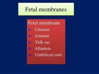
Fetal Tissues of the Fetal-Maternal Communication System • The extravillous and villous traphoblasts • Placental arm • The fetal membranes (the amnion-chorion leave) • Paracrine arm • Human placenta : hemochorioendothelial type
Early Human Development • Zygote • Blastomeres • Morula • Blastocyst • Embryo • Fetus • Conceptus
Fertilization of the Ovum and Cleavage of the Zygote • Moore, fig3-5
58-cell blastocyst • 107-cell blastocyst • Fig 5-1
Implantation • Moore, fig 3-4
Biology of trophoblast • Trophoblast is the most variable in structure, function and development • invasiveness provides for attatchment of blastocyst to decidua of uterine cavity • nutrition of the conceptus • function as endocrine organ in human pregnancy • essential to maternal physiological adaptations & maintenance of pregnancy
Differentiation • Cellular, syncytial/ uninuclear , multinuclear • Formation of the Syncytium
Cytotrophoblasts are the cellular progenitors of the syncytiotrophoblast
after apposition & adherence, intrusion of cytotrophoblast between endometrial epithelial cells • this process is facilitated by degradation of the extracellula matrix of endometrium /decidua catalyzed by • urokinase-type plasminogen activator • urokinase plasminogen activator receptor • multiple metalloproteinase • These functions of cytotrophoblasts invading the endometrium areindistinguishable from those of metastasizing malignant cells
Immunological Acceptance of the Conceptus • Previous Theories • antigenic immaturity of the embryo-fetus • diminished immunological responsiveness of the pregnant woman • Decidua : immunologically privileged tissue site • The acceptance and the survival of conceptus in the maternal uterus must be attributed to immunological peculiarity of the trophoblasts, not the decidua
Current Status of Research • Expression of the HLA system in trophoblast unique set of lymphocytes • > may provide explanation for immunological acceptance of the conceptus • 주로 trophoblast HLA expression (monomorphic HLA-G class I)과 uterine large granular lymphocyte (LGL)로 설명하고 있다. 그러나 아직은 완전하지 않다.
Immunocompetency of the Trophoblasts • Many researchers focused on the expression of the major histocompatibility complex (MHC) antigens in trophoblast • MHC class II antigens are absent from trophoblasts at all stages of gestation
Trophoblast HLA Class I Expression • Normal implantation is dependent upon controlled trophoblast invasion of maternal endometrium/decidua and the spiral arteries • a mechanism for permitting and then for limitting trophoblast invasion • Such a system involves the uterine large granular lymphocytes(LGSs) and the unique expression of specific nomomeric HLA class I antigens in the trophoblasts
HLA-I Gene Expression • HLA genes • the products of multiple genetic loci of the MHC within short arm of chromosome 6 • 17 class I genes have been identified • three classical genes • A, B, C => major class I(a) transplantation antigens • three other class I(b) genes • E, F, G => class I HLA antigen • HLA-G gene
Uterine Large Granular Lymphocyte (LGL) • Believed to be lymphoid and of bone marrow origin and natural killer cell lineage. • Present in large numbers only at the midluteal phase of the cycle-at the expected time of implantation in the human endometrium. • Near the end of luteal phase of nonfertile ovulatory cycles, the nuclei of LGLs beginto disintegrate. • With blastocyst implantation, these cells persist in the decidua during the early weeks of pregnancy. • speculated that LGLs are involved in the regulation of trophoblast invasion.
HLA-G Expression in Human Trophoblasts • HLA-G antigen • identified only in extravillous cytotrophoblast in decidua basails and chorion laeve • not present in villous trophoblast, either in syncytium or in cytotrophoblasts. • expressed in cytotrophoblast that are contiguous with maternal tissue (decidual cell) • It is hopothesized that HLA-G is immunologically permissive of antigen mismatch between mother and fetus.
HLA Expression in the Human Embryo • as gestation progresses, cells from inner cell mass of blastocyst gradually develop both class I and II HLA antigen • these tissuee are not in direct contact with maternal tissue or blood
Implantation and Integrin Switching • Apposition, adherence, then intrusion and invasion of the endometrium/decidua by cytotrophoblast(implantation) appears to be dependent upon • trophoblast elaboration of specific proteinases • degrade selected extracellular matrix proteins of the endometrium/decidua • coordinated and alternating process referred to as "integrin switching“ • facilitates migration and then attachment of trophoblasts in the decidua
Integrin • one of four families of cell adhesion molecules (CAMs) • cell-surface receptors that mediate the adhesion of cells to extracellular matrix proteins
Trophoblast Attachment in Decidua: Oncofetal Fibronectin • onfFN(oncofetal fibronectin) • unique glycopeptide of the trophouteronectin molecule • trophouteronectin or trophoblast glue • formed by extravillous trophoblast, including those of chorion laeve • Function • a critical role for migration and attachment of the trophoblasts to maternal decidua • facilitates separation of extraembryonic tissues from the uterus at delivery
Embryonic and Placental Development • Early Blastocyst • Trophoblast • hCG • Grow & expand
CytotrophoblastInvasion of DecidualVessels • Capillary network • arterioles • Spiral arteries
Several curious features • trophoblastsin the vessels lumen do not appear to replicate • these cells are not readily dislodged by flow of blood • these cytotrophoblastappear to migrate against arterial flow and pressure • no obvious adhesion of these cells one to the other • invasion of maternal vascular tissue bttrophoblastsinvolves only the decidual spiral arteries, not the veins
TrophoblastUltrastructure • Prominent microvilliof the syncytialsurface (brush border) • pinocytoticvacuolesand vesicles • absorptive and secretory placental function
ChorionicVilli • 12th day에 처음 발생 • Primary villi • proliferation of cytotrophoblastextend into syncytiotrophoblast • Secondary villi • mesenchymalcord, derived from cytotrophoblast, invade solid trophoblastcolumn • Tertiary villi • after angiogenesisoccurs from the mesenchymalcores in situ • 17th day에 fetal blood vessels are functional & placental circulation이 establish됨.
Characteristic of development of H-mole • some villi, in which absence of angiogenesisresults in a lack of circulation, may distended with fluid and form vesicles
Placental Cotyledons • Certain villiof the chorionfrondosumextend from chorionicplate to the decidua and serve as anchoring villi • Each of the main stem villi(truncal) and their ramifications (rami) constitute a placental cotyledon (lobe) • For each cotyledon, a 1:1:1 ratio of artery to vein to cotyledon
Breaks in the Placental " Barrier“ • Numerous findings of passage of cells between mother and fetus in both directions • ex) erythroblastosisfetalis • A few fetal blood cells are found in the mother's blood • Fetal leukocytesmay replicate in the mother and leukocytes bearing a Y chromosome have been identified in women for up to 5 years after giving birth to a son
PlacetalSize and Weight • Total number of cotyledonsremains the same throughout gestation • Individual cotyledonescontinue to grow • Placental weights vary considerably
Placental Aging • As villicontinue to branch and terminal ramifications become more numerous and smaller • > volume and prominence of cytotrophoblastsdecrease • As syncytiumthins and forms knots • > vessels become more prominent and lie closer to the surface • The stromaof the villi • in early pregnancy • branching connective ts. cells are seperatedby abundant loose intercellularmatrix • later • stromabecomes denser, and the cells more spindly and more closely packed
Histologicchanges that accompany placental growth and aging are suggestive of increase in the efficiency of transport to and exchange to meet increasing fetal metabolic requirements • decrease in thickness of the syncytium • partial reduction of cytotropholasticcell • decrease in the stroma • increase in the number of capillaries and approximation of these vessels to the syncytialsurface • By 4 months • the apparent continuity of the cytotrophoblastis broken • the syncytiumforms knots on the more numerous, smaller villi
At term • Covering of villimay be focally reduced to a thin layer of syncytium with minimal connective tissue • Fetal capillaries seem to abut the tropohoblast • Villousstroma, Hofbauercells, and cytotrophoblastsare markedly reduced • villiappear filled with thin-walled capillaries • Other changes suggestive of a decrease in the efficiency for placental exchange • thickening of the basement membrane of trophoblastcapillaries • obliteration of certain fetal vessels • fibrin deposition on the surface of villiin basal and chorionicplates as well as elsewhere in the intervillousspace
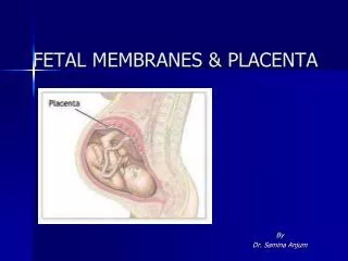
Blood Circulation in the Mature Placenta • A section through the placenta in situ • amnion → chorion→ chorionic villi → intervillous space → decidual plate → myometrium
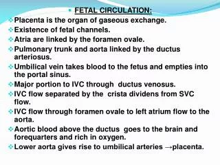
Fetal Circulation • 2 umbilical arteries • deoxygenated, or "venous-like" blood flows to the placenta • 1 umbilical vein • with a significantly higher oxygen content • Hyrtl anastomosis • Two umbilical a. separate at the chorionic plate to supply branches to the cotyledons
Maternal Circulation • Intervillous space -> chorionic plate -> vein • Spiral a. 는 수직으로, vein은 수평으로 주행 • Ut. Contraction하면 vein차단 • Intervillous space내에 정체되어 모체와 태아간의 물질교환 • Ramsey's concept
The principle factors regulating the flow of blood in the intervillous space • arterial blood pressure • intrauterine pressure • pattern of uterine contraction • factors that act specifically upon the arteriolar walls
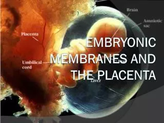
The Amnion • Innermost fetal membrane and is contiguous with amnionic fluid • Avascular structure • Provide almost all of the tensile strength of the fetal membranes • protect against rupture or tearing
Structure • single layer of cuboidal epithelial cells • basement membrane • acellular compact layer • fibroblast-like mesenchymal cells • zona spongiosa • Missing element of human amnion • smooth muscle cell, nerves, lymphatics, blood vessels