Placental US
Placental US. Andrea Jelks, MD 12/8/06. Case of the Day. 18 yo G2 P0101 at 18 weeks by 1 st trimester sono presented for an anatomy ultrasound Size = dates Fetus anatomically normal male Posterior placenta…. Outline. Various placental ultrasound findings Changes with gestational age

Placental US
E N D
Presentation Transcript
Placental US Andrea Jelks, MD 12/8/06
Case of the Day • 18 yo G2 P0101 at 18 weeks by 1st trimester sono presented for an anatomy ultrasound • Size = dates • Fetus anatomically normal male • Posterior placenta…
Outline • Various placental ultrasound findings • Changes with gestational age • Echolucencies Others…..
This image shows: • A posterior uterine contraction which gives a false impression of placenta previa • An anterior marginal placenta previa • A central placenta previa • A succenturiate lobe on the posterior uterine wall (A.) The internal os is poorly seen, but transvaginal US would almost certainly show that this placenta does not cover the cervix The thick area on the posterior uterine wall is a localized uterine contraction. The impression that there may be a previa is caused by the posterior uterine contraction. Source: www.obgyn.ufl.edu
Another picture of uterine contraction • Placenta would be of normal thickness if seen after the UC abated • Importance of reimaging after a few minutes have elapsed Source: Google images
This is a 21-week-fetus pregnancy c/b Rh sensitization. Hydrops with extensive edema, ascites, hydrothorax and abnormal thickness of placenta Placental thickness judged subjectively At midposition or cord insertion 2-4 cm = normal Source: www.thefetus.net These images show…
Normal Placental appearance • 8-20 weeks: uniform echotexture, 2-3 cm thickness • >20 weeks: • Measures 4-5 cm thick
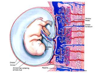
Decidua basalis Measures 9-10 mm thickness Contains maternal blood vessels Easy to confuse with retroplacental hemorrhage, especially if posterior placenta Fluid side = chorionic plate (chorioamniotic membrane) = bright specular reflector Source: www.mysono.com
Grade 0 Late 1st trimester-early 2nd trimester Uniform moderate echogenicity Smooth chorionic plate without indentations
Grade 1 Mid 2nd trimester –early 3rd trimester (~18-29 wks) Subtle indentations of chorionic plate Small, diffuse calcifications (hyperechoic) randomly dispersed in placenta
Grade 2 • Late 3rd trimester (~30 wks to delivery) • Larger indentations along chorionic plate • Larger calcifications in a “dot-dash” configuration along the basilar plate
Grade 3 39 wks – post dates Complete indentations of chorionic plate through to the basilar plate creating “cotyledons” (portions of placenta separated by the indentations) More irregular calcifications with significant shadowing May signify placental dysmaturity which can cause IUGR Associated with smoking, chronic hypertension, SLE, diabetes Found in ~20% of pregnancies at 40 weeks
Significance of Placental Grade • N = 1802 low-risk patients at 36 weeks • Grade III placenta found in 3.8% (68/1802). • Associated with young maternal age and cigarette smoking, p < 0.01. • PIH: Study group 7.4% (5/68) and 1.56% of controls (27/1734), p < 0.01. • SGA: 17.6% (12/68) vs. 5.6% (97/1734), p < 0.01. • Conclusion: Ultrasound detection of a grade III placenta at 36 weeks' gestation in a low-risk population helps to identify the "at-risk" pregnancy McKenna D, Ultrasonic evidence of placental calcification at 36 weeks' gestation: maternal and fetal outcomes. Acta Obstet Gynecol Scand. 2005 Jan;84(1):7-10.
This placenta would most likely be associated with: A) Fetal hydrops B) Trisomy 18 C) Lupus anticoagulant D) Maternal smoking (A.) This is a very thick, echogenic placenta in a fetus with hydrops. - Fetuses with Trisomy 18 have small placentas. - Pregnancies with lupus anticoagulant would have a small or normal placenta. - Maternal smoking results in an echogenic grade III placenta, but not diffuse thickening and echogenicity as seen here Source: www.obgyn.ufl.edu
All of the following statements are true except… • In the early second trimester approximately 5% of placentas appear to cover the internal os. • 80% of cases where previa is diagnosed in the early second trimester do not have placenta previa at term. • Since trophoblasts have the capacity to detach and reattach, many placentas migrate away from the internal os. • A full bladder can give a false positive diagnosis of placenta previa. • (C.) Trophoblasts do not have the capacity to detach and reattach. So called "migration" results from other factors. Source: www.obgyn.ufl.edu
This image demonstrates… • Placenta previa • A venous lake • An submembranous blood clot from an abruption • Amniotic band syndrome (C.) The appearance of this hypoechoic area between the membranes and uterine wall is very suggestive of a blood clot. The membranes are separated from the uterine wall. Amniotic band syndrome should not be diagnosed, however, unless there is evidence of attachments of the membrane to the fetus associated with fetal anomalies Source: www.obgyn.ufl.edu
Amniotic Bands • Multiple case reports of limb reduction/ deformities, facial clefts, and/or IUFD due to umbilical cord entanglement • Case controlled study (n = 25 cases vs. 50 controls) showed… • Unrestricted fetal movement on all US • No fetal abnormalities at birth • Increased incidence of PTD Wehbeh H et al. The relationship between the ultrasonographic diagnosis of innocent amniotic band development and pregnancy outcomes. Obstet Gynecol. 1993 Apr;81(4):565-8.
Which of the following is shown in this image? • A placental abruption • A grade III placenta • Venous lakes • Placental infarcts (D.) These hypoechoic areas surrounded by echogenic placenta are characteristic of infarcts. Infarcts frequently show an echogenic rim and absence of swirling blood on Doppler flow. Source: www.obgyn.ufl.edu
This area in the placenta represents: A) A large venous lakeB) An abruptionC) An infarctD) A chorioangioma This hypoechoic area on the surface of the placenta is characteristic of a venous lake. Blood flow can usually be seen with real time imaging with the gain set low. Represent pooling of maternal blood. An infarct would be a hypoechoic area within the placenta. An abruption would show a hypoechoic area below rather than on the surface of the placenta. Source: www.obgyn.ufl.edu
A rare placental tumor composed of vascular spaces. Usually seen as circumscribed solid mass or complex mass that protrudes from the fetal surface of the placenta. It has been postulated that these tumors begin around the 16th-17th day of development when a newly formed angioblastic mass becomes isolated from the rest of the proliferating trophoblast May cause… Cord compression AV fistula high output cardiac failure in the fetus hydrops Turbulence microangiopathic anemia hydrops Polyhydramnios is present in one third of the cases. Assoc with PTD, IUFD and IUGR Placental Chorioangioma Source: www.thefetus.net
These images show… Source: www.emedicine.com Source: Sharma et al, 2003
Clinical Significance of Subchorionic “Collections” • 122 cases of SCC in 10 years at 1 institution detected between 5-22 weeks, (no controls) • 63% c/b bleeding • 88 % of those with PTD had bleeding • 59 % of those del at term had bleeding • Outcomes: • 5 % SAB at 17-24 weeks • 18 % PTD (median 35 wks) • 77 % term delivery • Outcome not assoc. with maximal size, GA at detection Sharma, et al. Prognostic factors associated with antenatal subchorionic echolucencies. Am J Obstet Gynecol 2003;189: 994-6
Sharma, et al. Prognostic factors associated with antenatal subchorionic echolucencies. Am J Obstet Gynecol 2003;189: 994-6
Matched case control study 238 cases and 648 controls w/o VB Cases comprised 1.3% of total scanned population SAB: OR = 2.8 Stillbirth: OR = 4.5 Abruption: OR = 11.2 PTD: No difference Significance of 1st trim SCC Ball, RH, et al. The clinical significance of ultrasonographically detected subchorionic hemorrhages(Am J Obstet Gynecol 1996;174:996-1002.
Prospective study of those with 1st trimester hematoma (n=230) vs. controls (n=6488) Outcomes similar subchorionic vs. retroplacental location 18.7% had loss at less than 24 weeks Relative Risk (all signif.) Preeclampsia 4.0 Abruption 5.6 Retained Plac 3.2 IUGR 2.4 Fetal distress 2.6 Meconium 2.2 NICU admit 5.6 PTD NS Clinical Significance of 1st trim SCC Nagy et al. Clinical significance of subchorionic and retroplacental hematomas detected in the first trimester of pregnancy. Obstet Gynecol. 2003 Jul;102(1):94-100
This image shows….. • A marginal abruption • A large venous lake • A placental hemangioma. • A retroplacental abruption (D.) The hypoechoic area represents a blood clot behind the placenta. A placental hemangioma would be within the placenta, not behind the placenta as this mass is. This hypoechoic area underlies the placenta. Source: www.obgyn.ufl.edu
Placental abruption. This retroplacental hemorrhage is visualized in a patient during the third trimester of her pregnancy. Source: www.sunyabem.org/ultrasound.shtml
Nyberg et al.. Variety of ultrasonographic appearances Acute phase: hyperechoic to isoechoic 1 week: hypoechoic 2 weeks: sonolucent Glantz, et al… retrospective cohort study Sensitivity 24%, specificity 96%, and positive 88%, and negative predictive values 53% of ultrasonography for placental abruption. US diagnosis of Placental Abruption Oyelese Y, Ananth CV. Placental abruption. Obstet Gynecol. 2006 Oct;108(4):1005-16.
This image shows: • Placental abruption • Venous lakes • Partial mole • A degenerating fibroid (C.) The "Swiss-cheese" appearance of this placenta, with the presence of a fetus (seen to the right of the image) are characteristic of partial mole. A fibroid would have a more rounded appearance. The cystic spaces are within the placenta. These cystic spaces are scattered throughout the placenta. The irregular cystic areas are within the placenta itself, and don't have the appearance of a retroplacental blood clot Source: www.obgyn.ufl.edu
These images show… • Color Doppler scan in a 21-year-old woman in 33rd week of pregnancy (same patient as in Image 12) demonstrates prominent retroplacental vessels mimicking a retroplacental hematoma.
These images show… • Sagittal endovaginal scan of the uterus in a 29-year-old woman in 9th week of gestation demonstrates nonfusion and separation of chorion and amnion. • Source: www.emedicine.com/radio/topic662.htm
This image shows… • 6w3d, bleeding, dichorionic twins with one dead twin, subchorionic hemorrhage of both chorions, the living sac with a larger hemorrhage than the dead sac. • Source:www.obgyn.net/.../us/cotm/9904/cotm_9904
A few more details… • Per patient….Previous delivery at 28 weeks (baby weighed 1.5 lbs) after pt presented with sudden vaginal bleeding and contractions • No bleeding yet in this pregnancy