Aortic Emergencies
Aortic Emergencies. Moritz Haager Sept 19, 2002. Objectives. Define aortic dissection + AAA, and why its important Review pathophysiology and classification Discuss diagnostic modalities Discuss management options. Aortic dissection dissecting aortic aneurysm

Aortic Emergencies
E N D
Presentation Transcript
Aortic Emergencies Moritz Haager Sept 19, 2002
Objectives • Define aortic dissection + AAA, and why its important • Review pathophysiology and classification • Discuss diagnostic modalities • Discuss management options
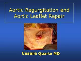
Aortic dissection dissecting aortic aneurysm “longitudinal cleavage of the aortic media created by a dissecting column of blood”Rosen’s Emergency Medicine What is Dissection?
Relevance • Underreported • Incidence 5-10/ 1,000,000 and rising • Mortality 1-2%/hour (33% in 1st 24 hrs) • High rate of misdiagnosis ~28% • One study suggests EP’s suspect AD in <50% of cases • Sullivan et al. Am J Emerg Med 18: 46-50. 2000 • Variable presentation including MI • Lack of suspicion for AD is #1 cause of misdiagnosis
Sad but true.. • 42 yo M presents c/o pain in R groin + leg • Also atypical CP/back pain preceding • Sniffed coke ~2 hrs ago + popped viagra ~1 hr ago at a “sex party” • PMHx: HTN, smoker • O/E: 92/ 160/100 /16; pale, diaphoretic, unwell looking; 2/6 SEM at apex, clear lungs, soft non-tender belly w/ Ø masses, R foot/ leg cool w/ Ø femoral, popliteal or tibial pulses • ECG: isolated ventricular beats • B/W: normal except +ve tox for cocaine • CT: Stanford Type B dissection • TEE: 3.5 cm intimal tear, Ø AR Famularo et al. J Emerg Med 21(1): 78-9. 2001
Requires 3 basic features: Abnormal media Blood entry into media (intimal tear, or vasa vasorum rupture) Pressure forces favoring propagation Pathophysiology
Pathophysiology • Peripheral complications: expanding hematoma dissects into, or compresses branches, or fistulizes into adj. structures • Cerebral CVA, syncope, • Spinal Neuro deficits • Cardiac MI, tamponade • Respiratory Hemoptysis, pleural effusion • GI Hematemesis, dysphagia, mesenteric ischemia • Renal ARF, HT N • Limbs Extremity ischemia
Intimal tears occur in 96% of all AD cases • Felt to occur 2o to shearing forces and hemodynamic stresses • Propagation factors: • Degree of HTN • Slope of pulse wave (dP/dT) • Spontaneous cure = rupture back into true lumen (rare)
Hemopericardium and tamponade can occur with dissection into pericardial sac
-Coronary artery involvement in ~1% presents as MI -0.1-0.2% of MI’s are complicated by admin of lytics in setting of AD
Clinical Presentation • Sudden, severe chest pain (~76-90%) • Migratory CP is highly specific (~71%) • Back pain (~53%), abd pain • Other Sx depending on site of involvement • Syncope (~9%) • Neuro Sx (~6-13%) • GI Sx • Resp Sx • Can be painless in up to 15% (chronic) Moore et al. Am J Card. 89:1235-1238 2002 Hals. Emerg Med Reports 2000
Risk Factors for AD • Hypertension (60-90%) • Age 50-70 yo • Male (3:1) • CTD’s (Marfans ~5%, Ehlers-Danlos) • Turners, Coarctation, Ebstein’s Anomaly • Congenital bi-/tricuspid AV • Family Hx or previous dissection • Cocaine, metamphetamine • Iatrogenic • Trauma Hals. Emerg Med Reports 2000
Classification • DeBakey: • Type I: involve ascending aorta, arch, and descending aorta • Type II: ascending aorta proximal to L subclavian artery • Type IIIa: descending aorta only; above diaphragm only • Type IIIb: descending aorta only; extension below diaphragm
Stanford • Type A: involvement of ascending aorta • Type B: no involvement of ascending aorta • ~62.5% of pts w/ AD have a Type A • Involvement of ascending aorta is of prognostic and therapeutic importance
How to diagnose AD • Clinical suspicion above all • 3 clinical variables shown to be useful: • Aortic pain (immediate onset, tearing, ripping) • Mediastinal widening / aortic widening on CXR • Pulse or BP differentials • Likelihood of AD: • Ø of above variables 7% risk of AD • Pain or widening alone 31 + 39% risk of AD respectively • > 2 variables or isolated BP / pulse diff > 83% risk of AD • Von Kodolitsch et al. Arch Intern Med. 160: 2977-82. 2000 • Diagnostic modalities • ECG, CXR, CT, TEE, Angiogram, MRI
ECG findings in AD • ~85% will be abnormal • LVH • Non-specific ST-T wave changes • MI (RCA most common) • Bottom line: • not sensitive or specific • beware thrombolysis until AD excluded
CXR findings in AD • 80-90% will be abnormal • Most findings non-specific: • Mediastinal widening (~59-75%) • Calcium sign (pathognomonic) • Double density aorta • Obliteration of aortic knuckle • Loss of PA window • Tracheal deviation to right • Depressed left main stem bronchus • New left pleural effusion • Apical cap • Size disparity of ascending + descending aorta
TEE • Rosen: 98% sensitive, 77% specific • Moore et al: 88% sensitive • 1st test in Europe and Japan • Advantages: • Can differentiate Type A + B dissections • Rapid, can be done at bedside • No contrast or radiation • Can detect AR and pericardial effusion • Disadvantages • Availability, operator dependence • Limited info on distal aorta Moore et al. Am J Card. 89:1235-1238. 2002
CT scanning • Dynamic helical CT nearly 100% sensitive and specific (dye) (Moore et al: 93% sens) • Advantages: • Availability, can differentiate Type A + B • Able to identify sealed-off false lumens • Able to identify other pathology (eg PE) • Disadvantages: • Dye reactions (1/10,000 fatal) • No info on AV function or intimal tear location • No info about extension into other arteries
Angiography • 81-87% sensitive, 96% specific • Previous gold standard • Advantages: • Anatomical delineation of aortic tree • Ability to demonstrate AR • Can differentiate Type A + B dissections • Disadvantages: • False –ves due to false lumen thrombosis • Invasive, time consuming, expensive
MRI • Near 100% sensitivity and specificity • No role in critical pts but good for serial follow-up • Advantages: • Excellent anatomical delineation, info on AR, intimal tear location, type and extent of AD • Disadvantages: • Time-consuming • Unable to monitor pt
Intravascular U/S • New technique – intravascular U/S probe • Currently evolving uses include • Identification of unstable plaques in CAD • Diagnostic and therapeutic use in AD • 3-D imaging of aorta + surrounding structures • Guidance of intra-vascular stent placement – less invasive procedure than classic surgery • Fenestration of intimal flap • Not available at most centers at this time Chavan et al. Circulation 96: 2124-2127. 1997
So what test do I order 1st? • Moore et al: CT is initial test of choice followed by TEE to clarify Dx or better delineate surrounding anatomy + AR • Moore et al. Am J Card. 89:1235-1238. 2002 • In Calgary, TEE is considered highly accurate and available, and should be considered a 1st line test • Peter Giannacarro, personal communication
Can we predict outcomes? • Pulse deficit is independent predictor of 5-day mortality RR 2.73, 95% CI 1.7-4.4 • Stat sig trend of increasing mortality with increasing number of pulse deficits • Bosssone et al. Am J Card 89: 851-855. 2002 • Mortality predictors: • Age > 70 (OR 1.7, 95% CI 1.05-2.77) • Abrupt onset CP (OR 2.6, 95% CI 1.22-5.54) • Hypotension/shock/tamponade (OR 2.97, 95% CI 1.25-3.29) • ARF (OR 1.77; 95% CI 1.80-12.6) • Pulse deficit (OR 2.03; 95% CI 1.25-3.29) • Abnormal ECG (OR 1.77; 95% CI 1.06-2.95) • Rajendra et al. Circulation. 105: 200-206. 2002
Management • ABC’s and then 2 basic principles: • Control dP/dT • 1st control your HR, then lower the pressure • Surgery if indicated • All Type A dissections need urgent OR • Controversies + New Ideas • Surgery for Type B dissections • Intravascular repair
Medical Tx • Control HR • IV BB’s – aim for HR 60-80 bpm • Propranolol 1mg IVP q5min • Esmolol 500 mcg/kg bolus, then titrate infusion 50-200 mcg/kg/min • Metoprolol 5 mg IVP q5min • Diltiazem 20 mg IV bolus, then 5-15 mg/hr if BB contraindicated (heart block, asthma, COPD, CHF)
Medical Tx • Control BP • aim for BP 100-120 mmHg sys or min BP req’d to maintain end-organ perfusion • Nitroprusside 0.5 mcg/kg/min – titrate up prn • Monotherapy • Labetalol 20 mg IVP, then 20-80 mg q5-10 min until in target HR, then 1-2 mg/hr
Surgical Tx • Indicated for: • All Type A dissections • Type B w/ complications: • Aortic rupture • Severe distal ischemia • Refractory HTN • Progressive dissection despite Tx • Intractable pain • Mortality for Type A repair is ~7-12% • Co-morbidities increase mortality • 5 yr survival is 77% Type A + 88% Type B
Why not operate on all? • Medical Tx of Type B has ~20% mortality • Surgical Tx of Type B has 10-15% mortality, and 3.5-36% risk of paraplegia • This may be changing with advent of endovascular repair, fenestration procedures, and IVUS.
New Surgical Methods • IVUS-guided fenestration of intimal flap • Chavan et al. Circulation 96: 2124-2127. 1997 • Intravascular stent placement • Cover intimal flap reducing flow to false lumen clotting of false lumen • 1.6 hrs vs 8 hrs for conventional Sx • One recent series of 70 Type B AD pts tx’d w/ stent-grafts reported 92.9% success and 9.6% all cause mortality at 29 months • Palma et al. Ann Thorac Surg 73: 138-42. 2002
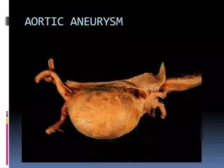
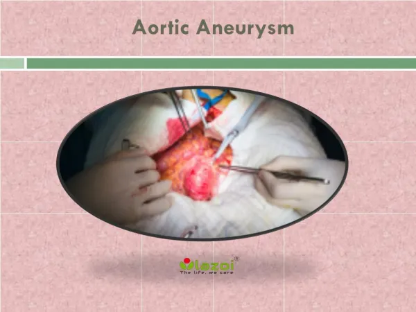
AAA: some facts • Incidence rising: 2% >65 yo • 9 men for every 1 female • Most have no antecedent Sx • 50-80% mortality rate • Misdiagnosed in 30-60% of cases
Definitions • Aneurysm = irreversible localized dilatation of an artery to > 1.5 original diameter (~3cm in abd aorta) • Types of aneurysms: • True = involves intima, media, and adventitia • Pseudoaneurysm = only intact + bulging layer is adventita • Inflammatory aneurysm = surrounding fibrosis and adhesions
Pseudoaneurysm • Damged intima + media • Adventitia prevents rupture
Anatomy • Aorta • Inf phrenic • R hepatic • Common hepatic • Gastroduodenal • Inf pancreatico-duodenal • L common iliac • L renal • Splenic • L gastric