Download
1 / 10
100 likes | 234 Vues
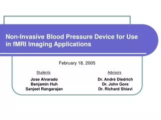
Non-Invasive Blood Pressure Device for Use in fMRI Imaging Applications. November 18, 2004. Students. Advisors. Jose Alvarado Ben Huh Sanjeet Rangarajan. Dr. Andr é Diedrich Dr. John Gore Dr. Richard Shiavi. Background.

E N D
Non-Invasive Blood Pressure Device for Use in fMRI Imaging Applications November 18, 2004 Students Advisors Jose Alvarado Ben Huh Sanjeet Rangarajan Dr. André Diedrich Dr. John Gore Dr. Richard Shiavi
Background • Functional Magnetic-Resonance Imaging (fMRI) has recently allowed novel insights into the function of individual brain sites. • Patients with baroreflex failure have extremely labile blood pressure due to loss of buffering function of blood pressure control. • Higher centers of the brain stem and cortical structures may have potentiating effects on changes in autonomic outflow. • Measuring blood pressure continuously during fMRI procedures could provide numerous benefits to the study of autonomic disorders.
The Problem • Current commercial devices such as the FINAPRES, FINOMETER, and PORTAPRES are able to continuously measure blood pressure but not in the presence of magnetic fields. • The electrical sensor system for the finger cuff as well as the pneumatic pump interfere with the highly sensitive fMRI magnet.
Market Analysis • Use for only in research and hospital settings. • Large market potential because of applications in other MRI and fMRI studies. • An optical continuous non-invasive blood pressure measuring device could be used in conjunction with electromagnetical trackers which are used in image guided surgery. • Profit will come from selling the design of the fMRI compatible finger cuff to pre-existing companies that manufacture the cuffs. • Perhaps too specific of an item to be able to market the device independently and for this reason it could be a better to sell the design to a current company.
Our Solution • Retrofit finger cuff blood pressure devices to use optical transmission techniques instead of electrical transmission techniques. • Replace electrical cables and sensors with optical components: • Fit cuff with an optical cable terminator. • Develop an fiber-optic interface compatible with existing commercially available electrical systems. • Design fMRI compatible shielding. • Extend length between cuff and electrical components without losing pneumatic function.
Completed Tasks • We have tested what we expected to be the first limiting factor, how far the pneumatic pump can be extended from the patient and still ensure proper cuff inflation. • Prediction: The small radius of the air tubes used with the FINAPRES creates a large resistance to flow with increasing tube length. Limited pump pressure leads to insufficient air supply to the cuff. • Experimental Result: Using tubing of the same diameter as the manufacturer, the maximum extension length was found to be around 10 ft.
Current Work • Currently we are testing tubing of varying radii and stiffness to determine the maximum length of extension while maintaining accurate blood pressure readings. • We are consulting with Dr. Gore about the possibility of shielding the electrical components in the case that an ideal extension length is unattainable using the manufacturer’s pneumatic pump.
Future Work • Contact Dr. Gore at the Vanderbilt University Institute for Imaging Science to become acclimated with the facility and learn about typical fMRI processes. The Imaging Institute’s fMRI scanner will eventually be used to test our design. • Dr. Duco Jansen will be contacted to ask for his expertise on selecting appropriate fiber-optic components. • Use experimental parameters to predict the necessary wavelength LED to ensure a proper signal and the photodetector needed to properly capture the transmitted light.
Questions Questions?