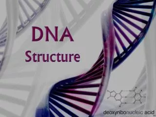
DNA STRUCTURE
DNA STRUCTURE. N UCLEIC ACIDS Include DNA: Deoxyribonucleic acid RNA: Ribonucleic acid. DNA ( Deoxyribonucleic acid): Site : Mostly found in the nucleus of cell and small amount is found in the mitochondria. DNA carries all the genetic informations of the individual

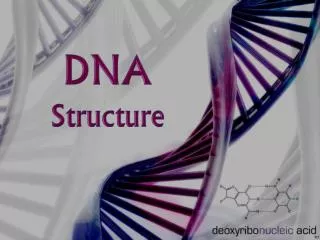
DNA STRUCTURE
E N D
Presentation Transcript
NUCLEIC ACIDS Include DNA: Deoxyribonucleic acid RNA: Ribonucleic acid
DNA (Deoxyribonucleic acid): • Site : Mostly found in the nucleus of cell and small amount is found in the mitochondria. • DNA carries all the genetic informations of the individual - Nuclear DNA:carries the genetic informations in the chromosomes that encode functional proteins or functional RNA. -
Mitochondrial DNA: • contains: • 1-genes encode proteins of the electron transport chain. • 2-genes encoding transfer RNA (tRNA) and the small and large subunits of ribosomal RNA (rRNA).
DNA must be to replicated precisely each time the cell divides, in such a way that each daughter cell acquires the same amount of genetic material.
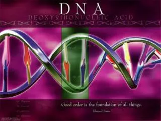
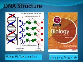
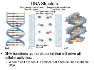
Structure: • DNA is formed of two chains (strands). • The 2 strands wind around each other forming a double helix (Watson and crick model).
Each strand (chain ) of DNA is formed of nucleotides • Each nucleotide of DNA is formed of : • Sugar (deoxy ribose). 2. Phosphate group. 3. Nitrogen containing bases: • Purine bases (bases containing 2 rings (Adenine [A], Guanine [G]) • Pyrimidine bases (bases containing single ring (Thymine [T], Cytosine)
Linking of the base to the sugar (at the 1’ carbon of the sugar) is called nucleoside Figure: β-Glycosidic linkage in a deoxyribonucleoside.
Linking of nucleoside to phosphate group ( at the 5’ carbon of the sugar) is called nucleotide
Nucleotides are linked in each DNA strand through Phosphodiester bond between the sugars and phosphates
Figure: Formation of a phosphodiester bond between 2 successive nucleotidesEster bonds are strong covalent bonds formed by the reaction of an acid and alcohol(- OH).Phosphodiester bond is a group of strong covalent bonds between a phosphate group and two 5-carbon ring carbohydrates (pentoses) over two ester bonds.
Fig.: A polynucleotide chain of DNA • Each DNA strand (chain) has 2 ends: • The 5’ end has a free phosphate group. • The 3’ end has a free hydroxyl group. • Note the direction of DNA replication is in a 5’ -3’ direction.
The hydrophilic (polar) deoxyribose phosphate backbone of each chain is on the outside of the DNA molecule, whereas the hydrophobic (nonpolar bases) are stacked inside perpendicular to the axis of the helix. • The 2 DNA strands wind around each other in antiparallel manner; that is From any fixed position in the helix, one strand is oriented in the 5′ 3′ direction and the other in the 3′ 5′ direction. • The double helix made by winding of the 2 DNA strands around each other, is stabilized by hydrogen bonding between the bases of the 2 strands and by the hydrophobic interactions between the stacked bases.
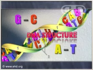
Base pairing rule: Thymine always pair with adenine and cytosine always pair with guanine. So one polynucleotide chain is of the double helix is always complementary to the other. • Thymine and adenine bases are connected by 2 hydrogen bonds , while cytosine and guanine are connected by 3 hydrogen bonds.This makes G-C base pairs more stable than A-T base pairs
On its exterior surface, the double helix of DNA contains two deep grooves between the ribose-phosphate chains. These two grooves are of unequal size and termed the major and minor grooves. The difference in their size is due to the asymmetry of the deoxyribose rings and the structurally distinct nature of the upper surface of a base-pair relative to the bottom surface.
The chromatin in eukaryotic cellsconsists of: Nuclear DNA bound to Histones proteins & smaller amounts of non histone proteins (Enzymes involved in DNA replication and transcription) & Small amount of RNA (snRNA)
The chromatin in eukaryotic cellsconsists of: Nuclear DNA bound to Histones proteins & smaller amounts of non histone proteins (Enzymes involved in DNA replication and transcription) & Small amount of RNA (snRNA)
HISTONES -Are basic proteins (having positive charges), so they form ionic bonds with the negatively charged DNA so their binding is facilitated. • - 5 types: H1, H2A, H2B, H3, H4. • - Functions: • They help condensation of the DNA into more compact chromosomes. • Protect the DNA from digestion by exonucleases. • Modified histones: Has an important role in changing the structure of chromatin and chromatin function e.g. acetylation of H3 and H4 is associated with activation or inactivation of gene expression.
Nucleosome consists of 1- DNA core: Formed of supercoiled DNA surrounding histone octamer(2 molecules of each of H2A, H2B, H3, H4). 2- Linking region: It joins one nucleosome core to the next Formed of 60 bp of DNA and 1 molecule of histone (H1) which protects the linker DNA from digestion by exonucleases
Chromatinis built from repeating nucleosomes units If the chromatin is placed in low salt buffer and viewed with E/M (i.e If the chromatin is pulled into a linear strucrure)→→→→ It resembles a string of beads( with the beads representing nucleosome cores and the string representing the DNA linker)
Compaction of chromatin • Condensation of the nucleosomes with histone H1 in the centreform the 10 nm fibril which represents 7 fold shortening of the linear ß- form DNA. • Supercoiling of the 10 nm fibril with 6-7 nucleosomes per turnform the 30 nm fiber which represents 50 fold shortening of the length of DNA. • Supercoiling of the 30 nm fiber intosuperloops(700 nm in diameter). • Each 6 superloops attached to a protein scaffold form a rosette. • Each 30 rosette form one coil. • Each 10 coils form one chromatid.
Chromosomes 2 identical sister chromatids attached at the centromere→→one chromosome (in the metaphase) The centromere - is rich in A=T and is about 130 bp long. - is connected to specific proteins to form a complex known as kinetochore which is connected to mitotic spindle. This complex is essential structure for chromosomal segregation during mitosis.