
Digestive System
Digestive System. Kelly Mitchell. Digestive System. This system is uniquely constructed to perform its specialized function by turning food into the energy we need to survive and prepares the residue for waste disposal . The roles of the Digestive System are to: take in food

Digestive System
E N D
Presentation Transcript
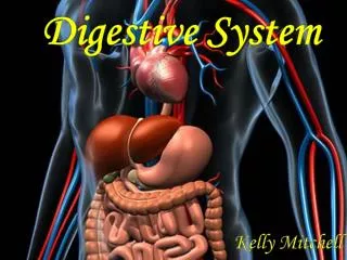
Digestive System Kelly Mitchell
Digestive System • This system is uniquely constructed to perform its specialized function by turning food into the energy we need to survive and prepares the residue for waste disposal. • The roles of the Digestive System are to: • take in food • break food down into nutrient • molecules • absorb molecules into the • bloodstream • rid the body of indigestible remains
Digestive System • The process of the Digestive System: (food becomes less complex at each process) • ingestion (taking in of food) • Propulsion (movement of food through the GI tract) • mechanical digestion (food is prepared for chemical ) • chemical digestion (complex food molecules are broken down into chemical building blocks) • Absorption (digested end products, vitamins, minerals and water are passed) • Defecation (indigestible substances are eliminated through the anus)
Digestive System • The organs of this system can be separated into two main groups: those forming the alimentary canal, and those forming the accessory digestive organs • The alimentary canal digests food, breaks it down into smaller fragments, and absorbs the digested fragments through its lining in the blood. • The accessory organs includes: the teeth, tongue, gall bladder, and digestive glands such as: the pancreas, liver, and salivary glands.
Alimentary Canal Organs • This is also called the gastrointestinal or the GI tract which is a continuous, coiled hollow muscular tube that winds through the ventral body cavity and has openings at both ends. • Its organs include: the mouth, pharynx, esophagus, stomach, small intestine, large intestine, and anus. • The essential activities of the GI tract are: • Ingestion • Propulsion • Food breakdown or mechanical digestion • Absorption • Defecation
What Organs make up the Digestive System? • The digestive tract consists of: • Mouth • Esophagus • Stomach • Small intestine • Large intestine (includes the colon and rectum) • Anus • Organs that Help with digestion: • Tongue • Pancreas • Liver • Gallbladder
Mouth • The mouth begins the digestive tract. • By chewing the food, this allows the body to be more easily digested. • The saliva in the mouth mixes with the food in your mouth and begins to break it down so your body can absorb the food. • The frenulum which is a fold of mucus membrane, secures the tongue to the floor of the mouth and limits its posterior movements. • The mouth in located in the center of the face directly below the nose.
Mouth • Food enters the digestive tract through the mouth, or oral cavity which is a mucous membrane lined cavity. • The lips protect its anterior opening, the cheeks form its lateral walls, the hard palate forms its anterior roof, and the uvula is a fleshy fingerlike projection of the soft palate, which extends downward from its posterior edge. • The area contained by the teeth is called the oral cavity proper.
Salivary Glands • Saliva is secreted by glands. • They are located in and outside the oral cavity. • The functions of the glands are: • cleanse the mouth • dissolve chemical food so it can be tasted • moisten food & compact it into a bolus • contain enzymes which begins the chemical digestion of starchy foods
Esophagus • This is located in your throat near your Trachea or windpipe. • Food travels to the Esophagus when it leaves your mouth. • This organ transfers food to your stomach by peristalsis or muscular contractions.
Stomach • The stomach is a hollow “container” which holds food while it is mixed with enzymes. • The enzymes continue to break down food into a form the body can use. • The cells in the lining of the stomach give out a strong acid and powerful enzymes. These aid in the breakdown process. • When the food in the stomach is completely processed, they are released into the small intestine.
Stomach • The activities of the stomach are food breakdown and food propulsion. • The stomach is a sac-like organ which has strong muscular walls. • The stomach is located in the lower abdominal cavity of the torso.
Small Intestine • The small intestine is made up of three parts: the duodenum, jejunum, and ileum. • This is a 22 foot long tube that uses enzymes to break down food. • The enzymes are released by the pancreas and bile from the liver. • Peristalsis or involuntary contractions move food and cause it to mix with digestive secretions from the liver and pancreas.
Small Intestine • The duodenum, the first part of the small intestine, is mostly responsible for the breaking-down process, and the jejunum and ileum are mainly responsible for nutrient absorption into the bloodstream. • The small intestine’s contents start out semi-solid, and end up as a liquid form after going through the small intestine.
Small Intestine • The change in consistency is caused by: water, bile, enzymes, and mucous. • Once the nutrients are absorbed and the leftover food liquid residue has passed through the small intestine, it then moves on to the colon or large intestine. • Activities of the small intestine are food breakdown and absorption and food propulsion.
Large Intestine, Colon • The Large Intestine is a 6-foot long muscular tube that connects the small intestine to the rectum. • This organ is made up of the cecum, the ascending (right) colon, the transverse (across) colon, the descending (left) colon, and the sigmoid colon, which also leads to the anus or terminal opening. • The appendix is a small tube which is attached to the cecum. • This organ is a highly specialized which is responsible for processing waste so emptying the bowels is convenient and easy.
Large Intestine, Colon • Waste left over from the digestive process is passed through the large intestine by peristalsis, first in a liquid state and ends up in a solid form. • As waste passes through the colon, water is removed. • Waste is stored in the sigmoid or the S-shaped colon until a mass movement empties it into the rectum once or twice a day. • This process normally takes about 36 hours for waste to get through the colon.
Large Intestine, Colon • The waste is made up of mostly food debris and bacteria. • These bacteria perform several useful functions such as: producing various vitamins, processing waste products, processing food particles, and protecting against harmful bacteria. • When the descending colon becomes full of waste it empties its contents into the rectum which begins the process of elimination. • Activities of the large intestine are food breakdown and absorption and propulsion of the residue and defecation or discharge from the anus.
Teeth • The teeth breaks down food in order to allow for easier digestion. • The Dental pulp’s function are: • connective tissue, blood vessels & nerve fibers • fills the pulp cavity enclosed by dentin • supplies nutrients to tooth tissues • provides for tooth sensation • The dentin in teeth is: • bone like material • underlies the enamel cap • forms the bulk of the tooth enamel • acellular, brittle, mineralized material • bears the force of chewing
Tongue • The muscular tongue occupies the floor of the mouth. • The tongue has several bony attachments two of which are to the hyoid bone and the styloid bone processes of the skull.
Pancreas • The pancreas secretes digestive enzymes into the duodenum which is the first segment of the small intestine. • The enzymes in the pancreas break down carbohydrates, fats, and, proteins. • The pancreas makes insulin, and secretes it directly into the bloodstream. • Insulin is the main hormone used for metabolizing sugar. • The pancreas is located in the lower cavity or the abdominal cavity.
Liver • The liver has many functions, but its main function in the digestive system is processing the nutrients absorbed in the small intestine. • Bile from the liver is secreted into the small intestine and plays an important role in the digestion of fat. • The liver is in a sense the body’s chemical "factory."
Liver • The liver takes the raw materials absorbed by the small intestine and makes all the chemicals the body needs to function. • This organ also removes potentially harmful chemicals. • It breaks down and gives off many drugs. • The liver is located in the lower cavity or the abdominal cavity.
Gallbladder • The gallbladder is a thin-walled, green sac. • It is also a ventral surface of liver. • The function of the gallbladder is to store bile that is not immediately needed for digestion. • The gallbladder also concentrates the bile by absorbing some of its water and ions.
Rectum • The rectum is an 8-inch chamber that connects the colon to the anus. • The rectum receives waste from the colon, lets the person know that there is stool to be evacuated, and holds the stool until evacuation happens. • When gas or waste comes into the rectum, sensors send a message to the brain.
Rectum • The brain decides if the waste contents are able to be released or not. • If they can be released, the sphincters or muscles relax and the rectum contracts and disposes of its contents. • If the contents can’t be disposed, the sphincter contracts and the rectum accommodates so that the sensation goes away temporarily.
Anus • The anus is the last part of the digestive tract. • It is a 2-inch long canal which consists of the pelvic floor muscles and the two anal sphincters both internal and external. • The lining of the upper anus is designed to detect rectal contents. • The anus lets you know whether the contents are liquid, gas, or solid. • The anus is surrounded by sphincter muscles that are important in allowing the control of stool.
Anus • The pelvic floor muscle creates an angle between the rectum and the anus which prevents stool from exiting when it is not supposed to. • The internal sphincter is always tight, except when stool comes into the rectum. • This organ keeps us content when we are asleep so we are unaware of the presence of stool. • When we get an urge to go to the bathroom, we rely on our external sphincter to hold the stool until reaching a toilet, where it then relaxes to release the waste.
Evolutionary Development • In amphioxus, which is any genus of lancelets, the digestive tract consists of only three parts: the oral cavity, the pharynx, and a tubular post pharyngeal, pharynx region, gut without subdivisions. The same condition holds in the most primitive living vertebrates such as lampreys and hagfishes. • In higher vertebrates the post pharyngeal gut is almost always subdivided into a series of regions that are both anatomically, part of the body, and functionally distinct. The most common is the esophagus, stomach, small intestine, large intestine, and rectum.
How to Maintain Homeostasis? • The integumentary system- provides nutrients needed by the skin. • Skeletal system- provides calcium and other nutrients for bone repair and growth. • Muscular system- provides glucose for muscle activity; the liver metabolizes lactic acid following anaerobic muscle activity. • Cardiovascular system- provides nutrients for plasma protein formation and blood cell formation; liver detoxifies blood, makes plasma proteins, destroys old red blood cells.
How to maintain homeostasis? • Lymphatic system- digestive tract provides nutrients for lmphatic organs; stomach acidity prevents pathogen invasion of the body • Respiratory system- breathing is possible through the mouth because the digestive and respiratory tract share the pharynx • Urinary system- liver synthesizes urea; digestive tract excretes bile pigments from the liver and provides nutrients
How to maintain Homeostasis? • Reproductive system- digestive tract provides nutrients for growth and repair of organs and for development of the fetus • Nervous system- digestive tract provides nutrients for growth, maintenance, and repair of neurons and neuorglia • Endocrine system- the stomach and small intestines produce hormones
How does the system communicate? • Communication to maintain homeostasis can occur through the nervous system or through chemical stimulation. One part of the nervous system controls the communication network that regulates bodily functions. This part of the nervous system functions without a person's thinking about it and without much noticeable indication that it is working. Chemicals used to communicate are called transmitters. Transmitters that are produced by one organ and travel to other organs through the bloodstream are called hormones. Transmitters that conduct messages between parts of the nervous system are called neurotransmitters. • One of the best known transmitters is the hormone adrenaline. Within moments, this chemical has the entire body on alert, a response sometimes called the fight-or-flight response. The heart beats more rapidly and powerfully, the eyes dilate to allow more light in, breathing quickens, and the activity of the digestive system decreases to allow more blood to go to the muscles. The effect is rapid and intense.
How does the system support cellular respiration? • Through the digestive system the body acquires the food it needs to fuel cells. The main food source needed are carbohydrates broken into glucose molecules. The cell breaks the glucose into something smaller and sends it off to the mitochondria. There the mitochondria uses these smaller pieces added to oxygen to make energy for you.
How does the arrangement of the organs determine the function of the system? • The arrangement of the organs allows the food to properly be prepared in the mouth then digested into the stomach and eventually expelled through the anus. The food must be broken down before it can travel down the esophagus and be churned in the stomach and expelled as waste through the anus.
What is feedback control? • Feedback control is a response within a system that influences the continued activity or productivity of that system. It is the control of a biological reaction by the end products of that reaction. It also helps maintain homeostasis in the human body.
What is thermoregulation? • Also called Heat Regulation, it is the maintenance of an optimum temperature range by an organism. Cold-blooded animals pick up or lose heat by way of the environment, moving from one place to another as necessary. Warm-blooded animals have additional means by which they can heat and cool their bodies. Muscular activity can be an important source of heat in both kinds of animals. In the same way humans must maintain how hot and cold they get and how much water the body contains.
Works Cited • Marieb, Elaine N. (2000). Essentials of Human Anatomy and Physiology. Holyoke, Massachusetts: Addison Wesley Longman, Inc. • Marshall, Karen. The Digestive System. Retrieved from http://www.montgomerycollege.edu/~kmarshal/BI205/ppt/chapt23-4.pdf. • human digestive system. (2013). In Encyclopedia Britannica. Retrieved from http://www.britannica.com/EBchecked/topic/1081754/human-digestive-system/45382/Evolutionary-development. • Maintenance of the Human Body: Digestive System and Nutrition. McGraw-Hill. Retrieved from http://highered.mcgraw-hill.com/sites/dl/free/0072347325/80938/mhb8ch05.pdf. • Na’auao, Ka Hana ‘Imi. (2009). Cellular Respiration. Ka Hana ‘ImiNa’auao. Retrieved from http://www.cds.hawaii.edu/kahana/downloads/curriculum/SectionII/Unit5/Unit5Appendix/5.X.B.CellularRespiration.pdf.
Works Cited • (2009). Disease and Conditions. The Cleveland Clinic Foundation. Retrieved from http://my.clevelandclinic.org/anatomy/digestive_system/hic_the_structure_and_function_of_the_digestive_system.aspx. • Beers MD, Mark H. (2006). Organ Systems. The Merck Manual Home Health Handbook. Retrieved from http://www.merckmanuals.com/home/fundamentals/the_human_body/organ_systems.html. • thermoregulation. (2013). In Encyclopedia Britannica. Retrieved from http://www.britannica.com/EBchecked/topic/591729/thermoregulation. • feedback. (2013). In Encyclopædia Britannica. Retrieved from http://www.britannica.com/EBchecked/topic/203684/feedback. • Kindersley, Dorling. (1994). Ultimate Visual Dictionary. New York, NY: DK Publishing Inc.
Picture Works Cited • human digestive system. (2013). In Encyclopedia Britannica. Retrieved from http://www.britannica.com/EBchecked/topic/1081754/human-digestive-system/45382/Evolutionary-development. • MotionCow. (2013). Digestive + Cardiovascular Systems. Turbo Squid. Retrieved from http://www.turbosquid.com/3d-models/human-digestive-3d-model/546475. • Tongue Cutting OK With Judge. (2009). J-Walk Blog. Retrieved from http://j-walkblog.com/index.php?/weblog/posts/tongue_cutting_ok_with_judge/. • Kene, Shubhangi. Stock Photo- Anatomy of a human stomach in blue background. 123RF. Retrieved from http://www.123rf.com/photo_14669746_anatomy-of-human-stomach-in-blue-background.html. • About Your Liver. Keck School of Medicine of USC. Retrieved from http://www.surgery.usc.edu/hepatobiliary/pg-aboutyourliver.html.


