Download
1 / 48
480 likes | 656 Vues
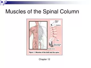
The Spinal Column and Thorax. ESAT 3600 Fundamentals of Athletic Training. Main Functions. Skull Protect brain Thorax Absorb forces of impact Muscle attachment Vertebral column Provides framework and foundation for most movements of body and extremities Muscle attachment

E N D
The Spinal Column and Thorax ESAT 3600 Fundamentals of Athletic Training
Main Functions • Skull • Protect brain • Thorax • Absorb forces of impact • Muscle attachment • Vertebral column • Provides framework and foundation for most movements of body and extremities • Muscle attachment • Protects spinal cord
Vertebral Column • 7 cervical • 12 thoracic • 5 lumbar • 5 sacral • 4-5 coccygeal • Sacral and coccygeal are fused
Shapes of Normal Spine • Cervical lordosis • Contributes to mobility of head • Thoracic kyphosis • Lumbar lordosis • Contributes to “springiness” of spine • Sacral kyphosis • Helps to transmit forces of body weight through trunk
Bony Anatomy Review • Vertebral body • Vertebral arch • Transverse processes • Spinous process • Articulating surfaces • facets
Vertebral Column Structure • All vertebrae are similar in general structure • 2 factors change when view from cervical to lumbar • Structures become progressively larger • Direction of articulating surfaces change
Facets • Different orientations allow for: • varied degrees of movement between 2 adjacent vertebrae • Change amounts of tension, compression, and shear forces
Vertebral Column Structure • Body of vertebra are load bearing portion of vertebra • Lumbar region bears more load than cervical region • Lumbar region has larger vertebrae
Cervical Vertebrae • Body is more rectangular shaped • Facets are flat and tilted anteriorly • Axis and atlas do not follow basic rules
Thoracic Vertebrae • Body has triangular shape • Facets are set oblique, and angle toward each other slightly
Lumbar Vertebrae • Body is rounder • Bean shaped • Facets are almost vertical and face each other
Intervertebral Disks • Consists of annulus fibrosis and nucleus pulposus
Annulus fibrosis • Outermost layer is regular fibrous tissue • Inner portion is fibrocartilage • Concentric layers
Nucleus Pulposus • Distributes stresses between vertebrae during flexion and extension • Lateral movements also • Forward bending = backward movement • Backward bending = forward movement
Disk Damage • Compression and shear of vertebral discs is associated with LBP • 3 factors that increase shear and compression • Amount of weight lifted • Distance held from the vertebral column • Amount of trunk flexion used when lifting
Ligaments of Vertebral Column • Anterior longitudinal ligament • Posterior longitudinal ligament
Ligaments of Vertebral Column • Supraspinous ligament • Interspinous ligament
Ligaments of Vertebral Column • Ligamentum flavum • Intertransverse ligament
Ligaments of Vertebral Column • Ligamentum nuchae • Atlantoaxial ligament • Transverse ligament • Hangman’s ligament • Transverse process to transverse process of atlas
Movement in the Spine • Movement between individual vertebrae • Movement of entire region • Movement of entire spine
Vertebral Column Stability • Weak bony stability • Most stability from ligaments and muscles • Muscle stabilizers • Abdominals • Prevent hyperextension • Erector spinae • Compresses column
Thorax • 12 Thoracic vertebrae • 12 pairs of ribs • 1st 7 have direct cartilaginous connection to sternum • Lower 5 connected to sternum by cartilaginous connection to the 7th costal cartilage or not at all
Trunk and Movement • Trunk helps to contribute force to movement • Movement of intervertebral joints position trunk so that the upper extremity is in optimal position • Throwing tasks
Spinal Muscles • Spinal muscle group • O & I on spinous process • Transversospinal muscle group • O on transverse process; I on spinous process • Spinotransversal muscle group • O on spinous process; I on transverse process
Role of Muscles in Trunk Alignment • Rectus abdominis • Prevents lumbar hyperextension & anterior pelvic tilt • Erector spinae group • Prevent lumbar flexion • Iliopsoas and rectus femoris • Prevent hip extension • Hamstrings and gluteals • Prevent hip flexion
Splenius capitis • O: spinous processes of C7-T3, inferior half of ligamentum nuchae • I: mastoid process and lateral 1/3 of superior nuchal line • A: bilaterally extends head and neck; unilaterally flexes and rotates head and neck to same side
Splenius cervicis • O: spinous process of 3rd-6th thoracic vertebrae • I: posterior transverse processes of the 1st, 2nd, 3rd cervical vertebrae, sometimes 4th cervical vertebrae • A: bilaterally extends head and neck; unilaterally laterally flexes and rotates neck to same side
Erector Spinae - Iliocostal Branch (Iliocostalis lumborum) • O: anterior surface of tendon arising from sacrum, spinous processes of lumbar and 11th & 12th thoracic vertebrae, medial lip of the iliac crest • I: inferior borders of angles of the lower 6 or 7 ribs • A: bilaterally extends spine; unilaterally lateral flexion of spine
Erector Spinae - Iliocostal Branch (Iliocostalis thoracis) • O: superior borders of the angles of the lower 6 ribs • I: into the angles of the upper 6 or 7 ribs and into the transverse process of the C7 • A: bilaterally extends spine; unilaterally lateral flexion of spine
Erector Spinae - Iliocostal Branch (Iliocostalis cervicis) • O: superior borders of the angles of the 3rd-6th ribs • I: posterior tubercles of transverse processes of the 4th, 5th, & 6th cervical vertebrae • A: bilaterally extends spine; unilaterally lateral flexion of spine
Erector spinae - Longissimus Branch (Longissimus thoracis) • O: common tendon w/ iliocostalis lumborum, fibers from transverse and accessory processes of lumbar vertebrae and thoracolumbar fascia • I: tips of transverse process of all thoracic vertebrae and the lower 9 or 10 ribs between the tubercles and angles • A: bilaterally extends vertebral column; draws ribs down; unilaterally laterally flexes vertebral column
Erector spinae - Longissimus Branch (Longissimus cervicis) • O: transverse processes of T 1-5 • I: transverse processes of C 2-6, sometimes transverse process of atlas • A: bilaterally extends neck; unilaterally laterally flexes neck
Erector spinae - Longissimus Branch (Longissimus capitis) • O: transverse processes of T 1-5, articular processes of C 4-7 • I: posterior margin of mastoid process • A: bilaterally extends head; unilaterally laterally flexes and rotates head to same side
Erector spinae - Spinalis Branch (Spinalis thoracis) • O: spinous processes of L 1-2, T11-12 • I: spinous processes of upper thoracic vertebrae • A: bilaterally extends spine; unilaterally laterally flexes spine
Erector spinae - Spinalis Branch (Spinalis cervicis) • O: lower portion of ligamentum nuchae, spinous processes of C7 and sometimes T1-2 • I: spinous processes of axis and sometimes spinous processes of C3-4 • A: bilaterally extends spine; unilaterally laterally flexes spine
Quadratus lumborum • O: iliolumbar ligament, posterior part of iliac crest • I: inferior border of 12th rib and transverse processes of upper 4 lumbar vertebrae • A: lateral flexion of lumbar spine, assist with inspiration
Multifidus • A series of pairs of small muscles extending the full length of the spine just superficial to the rotatores and each spanning 2 or 3 intervertebral spaces • O: posterior surface of sacrum, dorsal end of iliac crest, mamillary and transverse processes of L & T vertebrae, articular processes of C4-7 • I: spinous processes of all vertebrae except atlas • A: unilaterally laterally flexes and rotates spine to opposite side; bilaterally extends spine
Rotator brevis • Series of pairs of small muscles extending from sacrum to axis • O: transverse processes of vertebrae • I: bases of spinous processes of 1st vertebrae above • A: unilaterally rotation of spine to opposite side; bilaterally extends spine
Rotator longus • Series of pairs of small muscles extending from sacrum to axis • O: transverse processes of vertebrae • I: bases of the spinous processes of 2nd vertebrae above • A: unilaterally rotation of spine to opposite side; bilaterally extends spine
Scalenus anterior • O: scalene tubercle and ridge on superior surface of 1st rib • I: anterior tubercles of transverse of C3-6 • A: bilaterally flexes neck and raises 1st rib; unilaterally laterally flexes and rotates C spine to opposite side
Scalenus medius • O: superior surface of 1st rib behind subclavial groove • I: posterior tubercles of transverse processes of C2-7 • A: bilaterally flexes neck and raises 1st rib; unilaterally laterally flexes and rotates C spine to opposite side
Scalenus posterior • O: outer surface of 2nd rib behind attachment of serratus anterior • I: posterior tubercles of transverse processes of C4-6 • A: bilaterally flexes neck and raises 2nd rib; unilaterally laterally flexes and rotates C spine to opposite side
Sternocleidomastoid • O:Sternal head – anterior surface of manubrium; Clavicular head – superior surface of medial 1/3 of clavicle • I: lateral surface of mastoid process, lateral ½ of the superior nuchal line • A: bilaterally flexes head; unilaterally draws head to ipsilateral shoulder, rotates head to opposite side
External Oblique • O: external surfaces and inferior borders of ribs 5-12 • I: linea alba, inquinal ligament and anterior half of iliac crest along outer lip • A: unilaterally rotates trunk to opposite side, laterally flexes to same side; bilaterally flexes trunk anteriorly, anterior support to organs and lumbar spine, prevents anterior tilt of pelvis
Internal Oblique • O: lateral 2/3 of inquinal ligament and anterior 1/3 of middle line of iliac crest; middle 1/3 of iliac crest and thoraco lumbar fascia • I:crest of pubis and linea alba; inferior border of ribs 10-12 • A: unilaterally rotates and laterally flexes trunk to same side; bilaterally flexes spine
Rectus Abdominis • O: pubic crest and symphysis pubis • I: costal cartilage of ribs 5-7 and side of xiphoid process • A: flexion and lateral flexion of trunk, anterior support to organs and lumbar spine, prevents anterior tilt of pelvis
Transverse abdominis • O: lateral 1/3 of the inquinal ligament, anterior 2/3 of inner lip of iliac crest, thoracolumbar fascia, inner edges of lower 6 costal cartilages • I: linea alba • A: constricts abdominal contents, assists in forced expiration