Bone Marrow Collection Technique Sample Preparation Evaluation
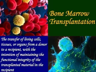
Bone Marrow Collection Technique Sample Preparation Evaluation. Clinical Pathology. Introduction to the hemopoietic system. Hematopoiesis : formation of blood cells The bone marrow is the major hematopoietic organ of the body.

Bone Marrow Collection Technique Sample Preparation Evaluation
E N D
Presentation Transcript
Bone MarrowCollection TechniqueSample PreparationEvaluation Clinical Pathology
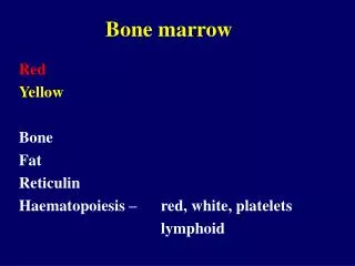
Introduction to the hemopoietic system • Hematopoiesis: formation of blood cells • The bone marrow is the major hematopoietic organ of the body. • In the adult (under normal circumstances), much of the bone marrow is hematopoietically inactive and filled with fat. • Active bone marrow remains in the flat bones and the ends of long bones. The central area has mainly fat. • Active bone marrow can expand into fat filled areas in response to increases peripheral use, loss, or destruction. • Remember red vs. yellow bone marrow? • In young animals, active hematopoietic tissue is found throughout both flat and long bones.
Indications for Bone Marrow Cytology • Hematologic abnormalities not readily explained by a good history, physical exam, chemistry panel, and/or other tests. • Nonregenerative anema • Persistent neutropenia • Persistent thrombocytopenia • Pancytopenia • Neoplasia • Proliferative disorders- myleofibrosis
Types of Bone Marrow Collection • Aspiration • Core biopsy
Problems with Bone Marrow Aspiration/Biopsy • Need to sedate or anesthetize patient • May be a risk • Hemorrhage • Concern with bleeding disorders or thrombocytopenic animals • Iatrogenic marrow infection • We cause an infection/issue
Common sites of collection of Bone Marrow Aspiration/Biopsy • Proximal end of the femur (trochanteric fossa) • Proximal end of the humerus • Iliac crest • Ribs • Sternum
Bone marrow aspiration/biopsy sites continued • Large dogs- iliac crest • Small dogs- trochanteric fossa of the femur • Cats- trochanteric fossa • The ribs and the sternum should be avoided in small dogs and cats- risk of puncturing the thoracic cavity • Biopsy of the trochanteric fossa may be difficult in obese or well-muscled animals
Instruments and Supplies • 16 to 18 gauge , 1 to 1 ¾ inch bone marrow biopsy needle. • 10-20 ml sterile syringe • Clean slides • Clear petri or watch glass • EDTA and saline • Surgery prep equipment • Sedative/anesthesia
Bone marrow collection technique • Sedate/anesthesize • Local anesthesia (in some cases) • Aseptic prep of area • Skin incision with blade • Biopsy needle is introduced and advanced into the cortical bone • Need is rotated in alternating clockwise and counterclockwise motions
Bone Marrow Collection Continued • Once in marrow cavity, the stylet is removed. • 10-20 ml syringe is attached and negative pressure is used to collect the marrow. • Apply suction until blood is seen within the hub of the syringe. • Stop at this point to avoid contamination with peripheral blood. • Place a drop of the sample onto clean slides.
Sample Preparation • Immediately place sample on a tilted slide • Allow the sample to drain from the slide into a watch glass or petri dish • Marrow flecks tend to adhere to the glass slide. • A second slide is placed perpendicularly across the marrow flecks causing it to spread • The 2 slides are then pulled apart in a horizontal plane • Marrow clots quickly so work quickly.
Using EDTA • An alternative is to use 3-5 ml of EDTA/isotonic saline in a syringe and then aspirate • Using this method will allow collection of a greater amount of sample and more slides may be made • The syringe contents are expelled into a watch glass or petri dish • The petri dish is tilted and/or rotated to examine the sample for marrow flecks • Marrow flecks are opaque/tan and irregular in shape. • Flecks cling to the bottom of the dish, the fluid drains to the bottom
Using EDTA continued • Harvest the flecks with microhematocrit capillary tube or pipette. • Place on slide, may need to blow gently over the top of the tube to dislodge the fleck • Using a coverslip use the horizontal pull apart technique. • Let air dry • Use Diff quick stain • Let stain and buffers allow a longer contact time
Core Biopsy technique • Same procedure except: • Jamshidi biopsy needle is used when the biopsy needle enters the marrow cavity, the stylet is removed and the needle is advanced about 3 mm with a rotating mtion. This cuts the core. • This fills the bore of the needle • The stylet or probe pushes the core out the top of the biopsy instrument • The core is rolled on the slide with the needle. • The remainder of the core is placed in formalin.
Bone Marrow Cells Types • Stem cells • Erythroid cells • Granulocytic cells • Monocytic cells • Megakaryocytic cells • Lymphocytic cells • Stromal and sustencacular cells (suppoting cells)
Stem Cells • Give rise to all blood cells depending on the body’s need • Erythroid series • Granulocytic series • Lymphocytic series • Monocytic series • Megakaryocytic series
Erythroid cells • Functions: to carry oxygen • The cells proliferate producing daughter cells • Remember Rubriblast to Reticulocyte
Granulocytic Cells • Functions depend on cell type • Neutrophils • Phagocytosis, mediators of inflammation, and microbiocidal actions • Eosinophils • Phagocytosis, parasiticidal, hyersensitivity reactions • Basophils • Inflammation and parasiticidal
Monocytic Cells • Function: • Tissue phagocytes (clean up functions), secrete mediators or inflammation, stimulate lymphocytes, and process antigens for presentation to lymphocytes
Megakaryocytic cells • Function: • Production of thrombocytes (important in hemostasis)
Lymphocytic cells • Function: • Mediation of the immune response (T-cells), antibody production (B-cells and plasma cells).
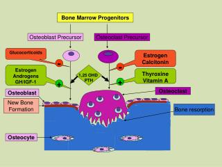
Other cells found in bone marrow • Vascular system cells • Supply nutrients to the marrow • Reticular cells • Give structure to the marrow • Osteoclasts and osteoblasts • Ocassionally found in an aspirate • Sometimes infectious organisms • Ehrlicia • Fungal • Leishmania
Basic pathologic lesions of the Bone Marrow • Hyperplasia • Hypoplasia • Neoplasia • Fibrosis • Inflammation • Infarction
Evaluation of the cellularity of the Marrow • Depends on the age of the animal • Young animals contain very little fat 25%, 75% cells • Adults contain 50% fat, 50% cells • Old animals contain 75% fat, 25% cells • Need to use other labwork to help differentiate the different causes of cellularity changes in the marrow • CBC • Chem panel • Clinical signs • History • Felv/FIV test • Ehrlichia, etc
Evaluating Bone Marrow Slides • Systematic approach • 10x scan slide- note degree of cellularity and amount of fat • Note the number of megakaryocytes • >50/large fleck suggests megakaryocyte hyperplasia • 80% of granulocytes should be more mature form • 90% of erythroid should be rubricytes and metarubricytes
Example of Bone Marrow Responses • Regenerative anemias tend to have hyperplastic erythroid compartment • Neutrophilia due to inflammation-hyperplastic with increased numbers of neutrophils • With Neutrophilia may see myeloid hypoplasia
Neoplastic Disorders • Leukemia is a neoplastic proliferation of hematopoietic cells within the bone marrow • Diagnosis is based on CBC and bone marrow exam • The bone marrow is replaced by proliferating immature cells • Lymphocytic leukemia • Lymphoblastic leukemia- more blast cells in the blood and bone marrow • Plasma cell myeloma- proliferation of plasma cells in the bone marrow.