Download
1 / 19
190 likes | 281 Vues
Explore the interaction between bacteria and biofunctionalized polymer-based nanoparticles in vitro and in vivo studies across various exposure routes. Results show no adverse cellular or tissue responses, supporting further research in nanoparticle design strategies. Acknowledging USDA and collaborators.

E N D
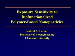
Exposure Sensitivity to Biofunctionalized Polymer-Based NanoparticlesRobert A. LatourProfessor of BioengineeringClemson University
Transmission electron micrograph of E. coli adhering to epithelium in the intestine of a pig. Moon, H.W. 1997. Comparative histopathology of intestinal infections. In: Mechanisms in the pathogenesis of enteric diseases (P.S. Paul, D.H. Francis and D.A. Benfield, eds.) Adv. Exptl. Med. Biol. 412:1. Plenum Press, New York.
Nanoparticles Bacterial cell High NP Concentration: Bacterial Isolation Intermediate NP Concentration: Bacterial Agglutination Low NP Concentration: Bacterial Tagging Bacterial Cell Binding Strategies
Mannose as Biofunctional group Polystyrene core Polyethylene glycol tether Nanoparticle Chemical Structure: Mannose Functionalization
Functionalized PEG side chains extending from hydrophobic polymer backbone chain. Diagram illustrates the self assembly into the nanoparticles followed by photochemical curing. Nanoparticle Design Strategy
a b c d e f E. coli - NP Interaction TEM images (dark-field) showing the agglutination of E. coli ORN178 mediated by D-mannose-tethered nanoparticles (a,b) Lower magnification and (c,d) higher magnification (e) E. coli ORN178only (similarly with bare nanoparticles) (f) E. coli ORN208 with the same D-mannose-tethered polymeric nanoparticles.
Acute Nanoparticle Exposure Sensitivity Studies • In vitro studies • cell toxicity studies • In vivo studies • Skin • Ocular • Inhalation • Ingestion • In vivo studies: poultry
In Vitro Results: Dermal Fibroblasts 1 ml cells + medium / 50 ml 2wt% np solution (core-PEG np) P = proliferating cells; NonP = nonproliferating cells np = with nanoparticles; C = control (w/o np)
Inhalation Study: Lung Tissue (fluorescence) 72 hr. Alveolar Sac / Alveolar duct nanoparticles Test (200x) Control (200x)
Inhalation study: Lung Tissue (H&E stain) Alveolar Sac / Alveolar duct Control (1000x) Test (1000x) Dark spots are nuclei of endothelial and connective tissue cells. Red spots are red blood cells. No detectable difference.
Oral Ingestion: Small Intestine Tissue (H&E stain) 72 hr. Transverse sections Away from center Towards center (Lumen) Control (400x) Test (400x) No apparent difference.
Oral Ingestion: Kidney (H&E stain) 72 hr. Control (400x) Test (400x) Glomerulus No apparent difference.
Oral Ingestion: Liver (H&E stain) 72 hr. Control (200x) Test (200x) No apparent difference.
Poultry Studies • 1-2 poults/pen gavaged with 0.1, 0.5 or 1.0 mL per • day of core-PEG nanoparticles, 2wt.%. • 3 control poults/pen gavaged with distilled water • Body weights at 1, 3 and 6 wk; observation to 14 wk • Commercial feed and water ad libitum
Poult Performance: 6-week Body Weight No significant effect of nanoparticles on poult body weight.
Concluding Remarks • In vitro & in vivo studies conducted with polystyrene-based nanoparticles. • No adverse cellular response for dermal fibroblast cells. • No apparent adverse tissue response from dermal, ocular, inhalation, or ingestion routes of exposure. • No adverse growth response from poultry studies. • Further in vitro and in vivo studies planned.
Acknowledgements • USDA for funding support • Collaborators: • Clemson University • F.J. Stutzenberger, T.-R.J. Tzeng, P.G. Luo, Dept. of Microbiology • Y.-P. Sun, L. Qu, S. Taylor, Dept. of Chemistry • S. Molugu, L. Jenkins, Dept. of Bioengineering • K. Bryant, J. Rodgers, Dept. of Envir. Toxicology • North Carolina State University, Dept. of Poultry Science • Jesse Grimes, B.W. Sheldon, J.L. Franklin, & M.J. Wineland