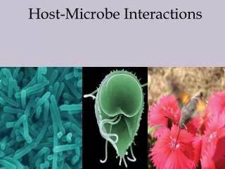
Bacterial Interactions with Host
Bacterial Interactions with Host. Medical Microbiology SBM 2044. When describing the interaction between host and microbes, analogies to war are common, as in references to a microbial “ attack ” or “ invasion ” repelled by host “ defense systems .”

Bacterial Interactions with Host
E N D
Presentation Transcript
Bacterial Interactions with Host Medical Microbiology SBM 2044
When describing the interaction between host and microbes, analogies to war are common, as in references to a microbial “attack” or “invasion” repelled by host “defense systems.” • In reality what drives microbes is notaggression but survival and reproduction. To succeed, many microbes have evolved the ability to persist in the body, only incidentally causing disease…
Pathogenesis • The pathogenesis of bacterial infection includes initiation of the infectious process and the mechanisms that lead to the development of signs and symptoms of disease. • Infectious process • Once in the body, bacteria must attach or adhere to host cells, usually epithelial cells • After establishment, the bacteria multiply and spread directly through tissues or via the lymphatic system to the bloodstream. • E.g. S. pneumoniae
Microbial survival • To survive, microbes must do the following: • Avoid being washed away (colonize the surfaces of host cells) • Find a nutritionally compatible niche • Survive the constitutive and induced defenses • Transfer to a new host
MCQ: True or False? • Microorganisms are the worst pathogens if they produce asymptomatic infection, rather than death of the host. (T/F ?) • Why? • False. Because pathogens that normally live in people enhance the possibility of transmission from one person to another.
Surface colonization • Once the bacteria enter the body of the host, they must adhere to cells or a tissue surface • avoid being wash away • compete with resident flora for adherence sites • Complex interactions • net surface charge • surface hydrophobicity • binding molecules on bacteria (ligands, adhesins) and host cell receptors
Overview of interactions with host surfaces • Nonspecific adhesion • Specific adhesion • overall, surface interactions • entrapment in mucin > 50 nm 10 – 20 nm < 2 nm < 1.0 nm Hydrophobic interactions Specific interactions Electrostatic repulsion Van der Walls Weak attractive adhesin receptor Weak long-range attractive easily disrupted Very strong irreversible Repulsion reduced by: (a) high ionic strength (b) small diameter
Nonspecific adhesion • Weakly adhering bacteria - easily removed by physical shear forces or washing • May allow colonisation of surfaces not subject to strong physical/washing forces (e.g. skin, vagina) • Not sufficient to colonise e.g. urinary tract, small intestine, etc
Specific adhesion EPEC adhering to an intestinal epithelial cells Bordetella pertussis on to ciliated tracheal cell
Specific adhesion The receptor specific molecule on host surface The adhesin specific molecule on bacterial cell surface Carbohydrate part of a glycolipid or glycoprotein Usually: A surface protein (often lectins) Others: e.g. LTA in Gram + • Specificity analogous to enzyme-substrate specificity
Types of bacterial adhesins • Individual protein molecules protruding from OM of Gram-neg. bacteria, or cell-wall of Gram-pos. bact. • protrude for distances ranging from < 10 - 20 nm, • up to ca.100nm (some Gram-pos. fibrillar adhesins) Example: Streptococcus pyogenes M proteins • Physical reasons why locating adhesins further away • from bacterial cell-surface – facilitate contacts with • receptors on mammalian cells
Bacterial Fimbriae • Adhesin often a minor subunit at tip • many 100’s of copies of major • structural subunit AFIMBRIAL adhesin • Assembly apparatus in OM • Specialized, multimeric, adhesive ‘appendages’ protruding several microns - much further than individual surface proteins. OM IM
Fimbriae on surface of a human ETEC strain • Strains may express > 1 distinct type of fimbriae, with different receptor specificities CS3 – thin, flexible CS1
Adhesin-Receptor specificity • First noted in late 1950s: • A particular soluble sugar, but not others, inhibited • adhesion of E. coli to cells (rbc adhesion model ) Specific adhesin Specific receptor • By mimicking critical part of the specific receptor, • inhibitor blocked receptor-binding site of adhesin • Type I pili: First E. coli adhesin identified • MSHA: mannose-sensitive haemagglutination
Other bacterial species • Wide variety of adhesins – both fimbrial & afimbrial (e.g. invasin that recognises integrins) • Some incorporate information on receptor-specificity in name: • Staphylococcus aureus: Fibronectin-binding proteins • Sialoprotein-binding protein • Streptococcus pyogenes: Fibronectin-binding proteins • Collagen-binding protein • Others first named for various reasons & only later discovered to act (also) as adhesins. • Streptococcus pyogenes: M proteins • Neisseria gonorrhoeae: Opa proteins
Consequences of adhesion Small intestine 1. Organism colonizes surface – e.g. normal flora 2. Pathogen colonizes surface and secretes toxins Examples: Vibrio cholerae Enterotoxigenic E. coli (ETEC) • Damage due mainly to action of secreted toxins • Damage often localized (e.g. cholera), but if toxin • absorbed efficiently (e.g. diphtheria), may be • systemic (i.e. throughout body)
Consequences of adhesion 3. Colonize surface and form a biofilm • In contrast to localised ‘colonies’, some pathogens can form a spreading surface layer – a ‘BIOFILM’ • bacteria encased in a polysaccharide slime that aids • attachment and protects bacteria. • ‘Simple’ biofilm: comprises a single species • Staphylococcus epidermidis • biofilm on a catheters
‘Complex’ Biofilms: • Comprise multiple species • Bacterial ‘co-aggregation’ • Some species produce polysaccharides, ‘trapping’ others Example: Dental plaque
Finding a Compatible Niche • Nutritious plasma contains sugars, vitamins and minerals… • but bacteria grow sparsely on fresh plasma in a test tube • plasma lack free iron • Bacteria adapt by: • use polymeric form of iron • siderophores which are specific for ferric iron • scavenging intracellular iron i.e. haemolysing • Specific nutrients only in certain body tissues • streptococci use sucrose in the mouth
Evading the Consitutive/Induced Defenses • The host possess • First line defense • Second line defense • How do microbial agents overcome these defenses?
Defending against complement • Microbes must prevent complement activation • Masking surface components (i.e. LPS, teichoic acids) that activate alternative pathway secreting capsules to cover up • Incorporate sialic acid into their capsular polysaccharides e.g. gonococci • Coating with circulating IgA – meningococci • Binding of C3b with viral envelope glycoprotein such as in herpes simplex virus • Prevent access of MAC to its target, the bacterial outer membrane – Salmonella and E. coli with long O-antigen polysaccharide chain
Avoiding phagocytosis • Many bacterial pathogens are rapidly killed once they are ingested by polymorphs or macrophages • But some pathogens are able to multiply within host cells, by shielding with normal host components on their surfaces
Avoiding phagocytosis • Inhibiting phagocyte recruitment and function • Bordetella pertussis inhibit neutrophil motility and chemotaxis – toxin production • Group A streptococci produce C5a peptidase – inactivates the chemotactic products
Avoiding phagocytosis • Microbial killing of phagocytes • Leukocidins kill neutrophils and macrophages – pseudomonads, staphylococci, group A streptococci, clostridia • shigellae kills phagocytic cells • Escaping ingestion • capsules • reduce opsonization e.g. staphylococci protein A binds to IgG by the wrong end, the Fc region
How microbes survive inside phagocytes? • Inhibition of lysosome fusion with phagosomes • Escapes into the cytoplasm • shigellae, Listeria monocytogenes and the rickettsiae cross the membrane of the phagocytic vesicle and the phagosome to enter cytoplasm, hence protected from lysozymes • L. monocytogenes secretes a pore-forming toxin, listeriolysin
How microbes survive inside phagocytes? • Resistance to lysosomal enzymes • Leishmania have resistant cell surfaces and can withstand acidic environment • Inhibition of the phagocytes ‘oxidative pathway’ • Legionella inhibit the hexose-monophosphate shunt and oxygen consumption in neutrophils, thus reducing the free radicals respiratory burst • staphylococci produce catalase that breaks down the H2O2
Intracellular life • Obligatory for viruses, but not bacteria. • Advantages? Protected from antibodies, and from some antimicrobial drugs • Intracellular microorganisms must be able to: • Penetrate • Survive host cell defenses • Transmit to other cells
Penetration • Easy with phagocytes • For non-phagocytic cells, penetration must be induced by microbial activities – • bind to specific receptors and send signals • Surviving host cell defenses • Transmission to other cells • upon lysis of the host cells, transmission through blood or body fluids • viruses spread by cell fusing with uninfected adjacent cells formation of syncytia and multinucleated giant cells • use of host cytoskeleton to spread e.g. Shigella, L. monocytogenes induce polymerization of actin at one of their ends
Subverting the immune responses • Immunosuppression • infectious agents may suppress the host’s immune responses. HIV infects the CD4+ T-helper lymphocytes which lead to the collapse of the immune system. • tuberculosis was more common during the measles outbreak • Diversion of lymphocyte function: superantigens • excessive stimulation of immune cells in a nonproductive way e.g. toxins secreted by streptococci are superantigens that cause misdirection of the immune response
Subverting the immune responses • Masquerading by changing antigenic coats • Trypanosomes • Trypanosoma brucei cause sleeping sickness • covered with thick protein coat called variable surface glycoprotein • have several hundred genes that encode various antigens, but only ONE is expressed at a time • Gonococci • periodic changes in pilin, protein of its pili • Influenza viruses • antigenic changes emerge gradually in a population of viruses over an epidemic season
Subverting the immune responses • Proteolysis of antibodies • extracellular proteases inactivate secretory IgA e.g. in gonococci, meningococci and Haemophilus influenzae • staphylokinase cleaves host plasminogen into plasmin at the bacterial cell surface • Viral latency • Herpes infection • herpes virus pass from one cell to another through cytoplasmic bridges • viruses do not proliferate within cells • other pathogens, Helicobacter pylori which causes gastric ulcers and Mycobacterium tuberculosis also maintain chronic associations with the host
Transmission of infection • Infectious agents are carried into a new host through food, aerosols, sexual contact, wound or soil • Many microbes differentiate into a transit form, to survive the environment • Chlamydiae – elementary body • Clinical manifestation of disease by microorganisms often promote transmission e.g. • Vibrio cholerae, E. coli and Shigella cause diarrhoea – intestinal contents secretion; • M. tuberculosis induce coughing –dispersal via aerosols