Download
1 / 29
410 likes | 1.07k Vues
Muscluar triangles of ……………….the neck. By: DR. AYAT ELDOMOUKY. POSTERIOR TRIANGLE. BOUNDARIES. Trapezius muscle. Sternomastoid muscle. clavicle. POSTERIOR TRIANGLE. Boundaries: - In front → Posterior border of sternomastoid m. - Behind → Anterior border of trapezius m.

E N D
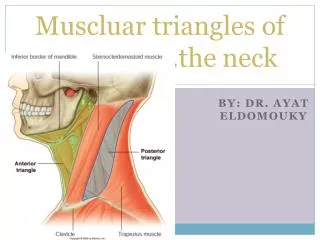
Muscluar triangles of ……………….the neck By: DR. AYAT ELDOMOUKY
POSTERIOR TRIANGLE BOUNDARIES Trapezius muscle Sternomastoid muscle clavicle
POSTERIOR TRIANGLE Boundaries: - In front → Posterior border of sternomastoid m. - Behind → Anterior border of trapezius m. - Apex → Middle ⅓ of superior nuchal line. - Base → Middle ⅓ of the clavicle. Roof - Skin. - Superficial fascia. - Deep (investing) fascia of the neck. The superficial fascia of the posterior triangle contains the following : (1) Platysma m. : (a subcutaneous m.) (2) External Jugular vein (3)The cutaneous branches of the cervical plexus Floor: From below upwards: a) Scalenusmedius. b) Levator scapulae. c) Splenius capitis.
Contents: Arteries 1- Third part of subclavian a. 2- Suprascapular a. 3- Superficial (or transverse) cervical a. 4- Occipital a. (near the apex). Veins 5- Subclavian v., receiving its only tributary, the external jugular v. Nerves 6- Roots and trunks of brachial plexus. 7- Spinal accessory n. (runs on levator scapulae). L.Ns. 8- Occipital L.N. (at the apex with the occipital a.). 9- Supraclavicular L.N. (at base with the subclavian a.). Ms. 10- Inferior belly of omohyoid .
DIVISION OF POSTERIOR TRIANGLE Occipital triangle Supra clavicular
ANTERIOR TRIANGLE □ Boundaries : • Anteriorly→ Middle line of the neck. • Posteriorly→ Ant. border of sternomastoid. • Base (above) → Lower border of mandible. • Apex (below) → Suprasternal notch. □ Roof: 1- Skin. 2- Superficial fascia (+ its contents). 3- Investing ( deep ) fascia. Contents of the Superficial Fascia of the Roof: a- Platysma muscle. b- Transverse cervical n. c- Anterior jugular v. : It is formed by the union of the submental veins → descends in the superficial fascia near the middle line → pierces the deep fascia above the sternum → passes deep to the sternomastoid → ends in the external jugular v. The deep parts of both anterior jugular veins are connected together by jugular arch. □ Subdivisions : 3 ½ triangles . See the table .
Suprahyoid muscles 1-MYLOHYOID MUSCLE Mylohyoid line
2- stylohyoid muscles 3- geniohyoid Inferior mental spine
Recall of relevant structures The suprahyoid muscles the muscles of the floor of the mouth
Enumerate main contents of submandibulartriangule and give short account on one of them Muscles: suprahyoid muscles (mylohyoid muscle), geniohyoid, stylohyoid, digastric and hyoglossus muscles Glands: submandibular salivary glands & L.N. Nerves: nerve to mylo-hyoid muscle, hypoglossal nerves and Submandibular ganglion Vessels: facial artery& vein
Enumerate main contents of submandibularregionand give short account on one of them Muscles: 1:suprahyoid muscles (mylohyoid muscle), geniohyoid, stylohyoid, digastric and hyoglossusmuscles 2:genioglossus Glands: submandibular salivary glands & L.N. Sublingual salivary gland Nerves: nerve to mylo-hyoid muscle, hypoglossal nerves and Submandibularganglion Lingual nerve Vessels: lingual & facial vessels
Facial vessels Submandibular ganglion Lingual nerve Sublingual salivary gland Hypoglossal nerve Lingual vessels Nerve to mylohyoid muscle Submandibular salivary gland
Enumerate main contents of carotid triangule and give short account on one of them Carotid sheath: CCA, ICA, IJV and vagus nerve External carotid artery Hypoglossal nerve Ansacervicalis Accessory nerve Sympathetic trunk
ANSA CERVICALIS • It is a nervous loop situated in front of the carotid sheath. • It is formed by union of 2 limbs : A) Superior limb : Descending from the hypoglossal n. and containing fibers of C1 spinal nerve . B) Inferior limb : From C2 and C3 spinal nerves . • Branches: arise from the tip → supply all infrahyoid muscles (near their lower ends) except thyrohyoid.
Enumerate main contents of muscular triangule and give short account on one of them Infra hyoid muscles
Enumerate main contents of submentaltriangule and give short account on one of them Submental LN Anterior jugular vein