Download
1 / 51
900 likes | 5.74k Vues
Bleeding disorders in children. Abdulhadi Alzaben , MD Pediatric hematology oncology and stem cell transplant. hematopoiesis.

E N D
Bleeding disorders in children AbdulhadiAlzaben, MD Pediatric hematology oncology and stem cell transplant
hematopoiesis • Hematopoiesis begins by 3 weeks of gestation with erythropoiesis in the yolk sac. By 2 months’ gestation, the primary site of hematopoiesis migrates to the liver. By 5 to 6 months’ gestation, the process shifts from the liver to the bone marrow. • During infancy, virtually all marrow cavities are actively hematopoietic and the proportion of hematopoietic to stromal elements is quite high. • As the child grows, hematopoiesis moves to the central bones of the body (vertebrae, sternum, ribs, and pelvis), and the marrow is gradually replaced with fat.
The normally high hemoglobin level of the fetus is a result of fetal erythropoietin production in the liver in response to low Po2 in utero. • Normal life span of The RBCisabout 120 days. • Embryonic hemoglobin's are produced during yolk sac erythropoiesis, then replaced by fetal hemoglobin (hemoglobin F, α2γ2) during the hepatic phase. • During the third trimester, gamma chain production gradually diminishes, replaced by beta chains, resulting in hemoglobin A (α2β2).
During the first few months of postnatal life, rapid growth, shortened RBC survival, and cessation of erythropoiesis cause a gradual decline in hemoglobin levels, with a nadir at 8 to 10 weeks of life. • This so-called physiologic nadir is accentuated in premature infants. • By 6 months of age in healthy infants, only trace gamma chain synthesis occurs.
Production of neutrophil precursors is controlled predominantly by two different colony-stimulating factors. • The most immature neutrophil precursors are controlled by granulocyte-macrophage colony-stimulating factor (GM-CSF), produced by monocytes and lymphocytes. • Granulocyte colony-stimulating factor (G-CSF) augments the production of more mature granulocyte precursors. • The rapid increase in neutrophil count that occurs with infection is caused by release of stored neutrophils from the bone marrow, under the control of GM-CSF.
Neutrophils migrate from the bone marrow, circulate for 6 to 7 hours, and enter the tissues, where they become end-stage cells that do not recirculate. • Eosinophil production is under the control of a related glycoprotein hormone, interleukin 3. Eosinophils, which play a role in host defense against parasites, also are capable of living in tissues for prolonged periods. • The bone marrow is the major storage organ for mature neutrophils and contains about seven times the intravascular pool of neutrophils. • It contains 2.5 to 5 times as many cells of myeloid lineage as cells of erythroid lineage. Smaller numbers of megakaryocytes, plasma cells, histiocytes, lymphocytes, and stromal cells are also stored in the marrow.
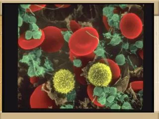
Megakaryocytes are giant, multinucleated cells derived from the primitive stem cell and are polyploid (16 to 32 times the normal DNA content). • Thrombopoietin is the primary regulator of platelet production. Platelets adhere to damaged endothelium and subendothelial surfaces via specific receptors for the adhesive proteins, von Willebrand factor (vWF), and fibrinogen. • Platelets also have specific granules that readily release their contents after stimulation and trigger the process of platelet aggregation. • Platelets circulate for 7 to 10 days and have no nucleus.
Functional Classification of Bleeding Disorders • Abnormal vessels Ehler Danlos HSP • Defects of platelets Number: Infection, ITP, Leukemia Function: vWD, BS, GT, Drugs • Abnormal coagulation Congenial: Hemophilia A or B Acquired: Liver disease, Vit K def, DIC
History: Neonatal bleeding Bleeding after circumcision Delayed bleeding from umbilical stump (factor XIII) Deep hematoma after I.M injections Epistaxis Unilateral vs bilateral Tooth extraction, tonsils Site of bleeding: skin, mm, joints Family history of bleeding disorders Drug exposure Injury child abuse
A male infant who is starting to walk and presents with a painful swollen joint after a fall has hemophilia until proven otherwise. • An adolescent girl who presents with excessive menstrual bleeding, recurrent nosebleeds, and pallor may have von Willebrand Disease (vWD), the most common inherited bleeding disorder • A five-year-old child who is not clinically ill but presents with moderate mucocutaneous purpura in the wake of a viral infection most likely has acute post-infectious immune thrombocytopenia • A teenage girl with easy bruising and mild pallor presenting to a pediatrician's office with a strong family history of autoimmune disorders may have chronic ITP • A ten-day-old infant with bleeding from the umbilical stump should be evaluated for factor XIII deficiency ,Intracranial hemorrhage in an infant without other risk factors should also prompt consideration of this diagnosis.
Physical examination: Petichiae Ecchymosis Joint bleeding and deep seated hematomas Significant lymphadenopathy Hepatosplenomegaly Active and playful vs ill looking Telangietic vessels Hemangiomas Loose joints and lax skin associated with easy bruising (Ehlers-Danlos syndrome)
Diagnosis of Coagulation Disorders Screening Tests • Complete blood count : • Thrombocytopenia when less than 150,000/mm3 • Platelet count is essential in the evaluation of the child with a positive bleeding history because thrombocytopenia is the most common acquired cause of a bleeding diathesis in children. • PseudothrombocytopeniaExamination of the peripheral blood smear is essential in patients with low platelet counts in order to exclude the presence of pseudothrombocytopenia caused by platelet aggregation after using EDTA as an in vitro anticoagulant
Prothrombin time (PT) : • The production of fibrin via the extrinsic pathway and the final common pathway requires tissue thromboplastin (tissue factor), factor VII, factors X, V, prothrombin (factor II), and fibrinogen. • This test bypasses the intrinsic pathway and uses "complete" thromboplastins (ie, tissue factor) capable of activating the extrinsic pathway. • The PT is sensitive to alterations in the vitamin K-dependent coagulation factors, especially factors II, VII, and X, and is used to monitor treatment with vitamin K antagonists In most laboratories, the normal PT value is 10-13 sec. PT has been standardized using the international normalized ratio (INR) so that values can be compared from one • laboratory or instrument to another. • This ratio is used to determine similar degrees of anticoagulation with warfarin (Coumadin)–like medications.
Activated partial thromboplastin time (aPTT) • The aPTT measures the intrinsic and common pathways of coagulation • This aPTT is routinely used to evaluate intrinsic coagulation and the degree of heparinanticoagulation. • The aPTT is sensitive to deficiencies of factors XII, XI, IX, and VIII and to inhibitors such as heparin • It is less sensitive than the PT to deficiencies within the common pathway (eg, factors X, V, prothrombin, and fibrinogen) and is unaffected by alterations in factors VII and XIII.
Thrombin Time: • Thrombin time measures the final step in the clotting cascade, in which fibrinogen is converted to fibrin. • The normal thrombin time varies between laboratories but is usually 11-15 sec. • Prolongation of thrombin time occurs with reduced fibrinogen levels (hypofibrinogenemia or afibrinogenemia), and dysfunctional fibrinogen (dysfibrinogenemia)
Bleeding Time: • Bleeding time assesses the function of platelets and their interaction with the vascular wall • Bleeding time is a difficult laboratory test to standardize • Platelet Function Analyzer: • evaluate the early stages of hemostasis (platelet function and VWF interaction under high shear • The PFA-100 measures platelet adhesion -aggregation in whole blood at high shear when exposed to either collagen-epinephrine or collagen-ADP. • Results are reported as the closure time measured in sec. • Clotting Factor Assays:
Disorders of Platelets • Platelet counts less than 150,000/mm3 constitute thrombocytopenia. Mucocutaneous bleeding is the hallmark of platelet disorders • Children with platelet counts less than 20,000/mm3 are at risk for spontaneous bleeding. • The etiology of thrombocytopenia may be organized into two mechanisms: 1 Decreased platelet production 2 Increased destruction
Decreased Platelet Production: • Thrombocytopenia with absent radii syndrome (TAR) is characterized by severe thrombocytopenia in association with orthopedic abnormalities, especially of the upper extremity. The thrombocytopenia usually improves over time • Congenital amegakaryocytic thrombocytopenia(CAMT) presents at birth or shortly thereafter with findings of severe thrombocytopenia, but no other congenital anomalies. The marrow is devoid of megakaryocytes and usually progresses to aplasia of all hematopoietic cell lines. • Acquired thrombocytopenia as a result of decreased production is rarely an isolated finding. It is seen more often in the context of pancytopenia resulting from bone marrow failure caused by infiltrative or aplastic processes.
Peripheral Destruction: ImmuneThrombocytopenic Purpura • Autoimmune thrombocytopenic purpura of childhood (childhood ITP) is a common disorder that usually follows an acute viral infection. Childhood ITP is caused by an antibody (IgG or IgM) that binds to the platelet membrane. • Clinical Manifestations. Young children typically exhibit ITP 1 to 4 weeks after viral illness, with abrupt onset of petechiae, purpura, and epistaxis. • The thrombocytopenia usually is severe. Significant adenopathy or hepatosplenomegaly is unusual, and the red blood cell (RBC) and white blood cell (WBC) counts are normal.
The diagnosis of ITP usually is based on clinical presentation and the platelet count and does not often require a bone marrow examination. • If atypical findings are noted, however, marrow examination is indicated to rule out an infiltrative disorder (leukemia) or an aplastic process (aplastic anemia). • BM study before steroid treatment • In ITP, an examination of the bone marrow reveals increased megakaryocytes and normal erythroid and myeloid elements. • Therapy is seldom indicated for platelet counts greater than 30,000/mm3. • Therapy does not affect the long-term outcome of ITP but is intended to increase the platelet count acutely.
For moderate and severe clinical bleeding with severe thrombocytopenia (platelet count <10,000/mm3), therapeutic options include prednisone, 2 to 4 mg/kg/24 hours for 2 weeks or IVIG, 1 g/kg/24 hours for 1 to 2 days. • Splenectomy is indicated in acute ITP only for life-threatening bleeding. • Approximately 80% of children have a spontaneous resolution of ITP within 6 months after diagnosis. • Serious bleeding, especially intracranial bleeding, occurs in fewer than 1% of patients with ITP. There is no evidence that early treatment prevents intracranial bleeding.
ITP that persists for 6 to 12 months is classified as chronic ITP. • Repeated treatments with IVIG, IV anti-D, or high-dose pulse steroids are effective in delaying the need for splenectomy. • Rituximab ( anti CD20) induces remission in 50% of cases • Secondary causes of chronic ITP, especially SLE and HIV infection, should be ruled out. • Splenectomy induces a remission in 70% to 80% of childhood chronic ITP cases. • The risks of splenectomy (surgery, sepsis from encapsulated bacteria) must be weighed against the risk of severe bleeding.
Wiskott-Aldrich syndrome • Is an X-linked disorder characterized by hypogammaglobinemia, eczema, and thrombocytopenia • Small platelets are seen on a peripheral blood smear. • Hemolytic uremic syndrome occurs as a result of exposure to a toxin that induces endothelial injury, fibrin deposition, and platelet activation and clearance • Thrombotic thrombocytopenic purpuraplatelet consumption, precipitated by a congenital or acquired deficiency of a metalloproteinase that cleaves von Willebrand factor, seems to be the primary process, with a modest deposition of fibrin and RBC destruction.
Disorders of Platelet Function • Primary disorders of platelet function may involve receptors on platelet membranes for adhesive proteins. • Deficiency of glycoprotein Ib complex (vWF receptor) causes Bernard-Soulier syndrome. • A deficiency of glycoprotein IIb- IIIa (the fibrinogen receptor) causes Glanzmannthrombasthenia.
Secondary disorders caused by toxins and drugs (uremia, valproic acid, aspirin, nonsteroidal anti-inflammatory drugs, and infections) may cause a broad spectrum of platelet dysfunction. • Disorders of platelet function present with mucocutaneous bleeding and a prolonged bleeding time or long PFA closure time and may be primary or secondary.
Disorders of Clotting Factors • The genes for factor 8 and factor 9 are on the X chromosome, whereas virtually all the other clotting factors are coded on autosomal chromosomes. • Factor 8 and factor 9 deficiencies are the most common severe inherited bleeding disorders. • von Willebrand disease is the most common congenital bleeding disorder. • factor 12 cause a prolonged activated partial thromboplastin time but are not associated with a predisposition to bleeding.
Hemophilia A (factor 8 deficiency) occurs in 1 in 5000 males. • Hemophilia B (factor 9 deficiency) occurs in approximately 1 in 25,000. • Clinically the two disorders are indistinguishable other than by their therapy • The severity of the disorder is determined by the degree of clotting factor deficiency.
Patients with less than 1% (severe hemophilia) factor 8 or factor 9 may have spontaneous bleeding or bleeding with minor trauma. • Patients with 1% to 5% (moderate hemophilia) factor 8 or factor 9 usually require moderate trauma to induce bleeding episodes. • In mild hemophilia (>5% factor 8 or factor 9), significant trauma is necessary to induce bleeding; spontaneous bleeding does not occur.
Mild hemophilia may go undiagnosed for many years, whereas severe hemophilia manifests in infancy when the child reaches the toddler stage. • In severe hemophilia, spontaneous bleeding occurs, usually in the muscles or joints • The diagnosis of hemophilia is based on a prolonged activated partial thromboplastin time (aPTT).
When an abnormal aPTT is obtained, specific factor assays are needed to make a precise diagnosis to determine the appropriate factor replacement therapy. • Early, appropriate replacement therapy is the hallmark of excellent hemophilia care. • Acute bleeding episodes are best treated in the home when the patient has attained the appropriate age and the parents have learned home treatment.
Bleeding associated with surgery, trauma, or dental extraction often can be anticipated, and excessive bleeding can be prevented with appropriate replacement therapy. • Prophylactic therapy starting in infancy has greatly diminished the likelihood of chronic arthropathy in children with hemophilia. • For life-threatening bleeding, levels of 80% to 100% of normal factor 8 or factor 9 are necessary.
For mild to moderate bleeding episodes (hemarthroses), a 40% level for factor 8 or a 30% to 40% level for factor 9 is appropriate. • The dose can be calculated using the knowledge that 1 U/kg body weight of factor 8 increases the plasma level 2%, whereas 1.5 U/kg of recombinant factor 9 increases the plasma level 1% • Desmopressin acetate is a synthetic vasopressin analog with minimal vasopressor effect. Desmopressin triples or quadruples the initial factor 8 level of a patient with mild or moderate (not severe) hemophilia A, but has no effect on factor 9 levels
desmopressin is the treatment of choice for individuals with mild and moderate hemophilia A. • Aminocaproic acid is an inhibitor of fibrinolysis that may be useful for oral bleeding. • Patients treated with older factor 8 or 9 concentrates derived from large pools of plasma donors were at high risk for hepatitis B, C, and D and HIV. • Recombinant factor 8 and factor 9 concentrates are safe from virally transmitted illnesses.
Inhibitors are IgG antibodies directed against transfused factor 8 or factor 9 in congenitally deficient patients. Inhibitors arise in 15% of severe factor 8 hemophiliacs but are less common in factor 9 hemophiliacs. • The treatment of bleeding patients with an inhibitor is difficult. For low titer inhibitors, options include continuous factor 8 infusions. For high titer inhibitors, it is usually necessary to administer a product that bypasses the inhibitor, preferably recombinant factor 7a. • Early institution of factor replacement and continuous prophylaxis beginning in early childhood should prevent the chronic joint disease associated with hemophilia
von Willebrand Disease • Von Willebrand disease is a common disorder (1% of the population) caused by a deficiency of vWF, an adhesive protein that serves two functions: • acting as a bridge between subendothelial collagen and platelets and binding and protecting circulating factor 8 from rapid clearance from circulation. • Von Willebrand disease usually is inherited as an autosomal dominant trait and rarely as an autosomal recessive trait.
Approximately 80% of patients with von Willebrand disease have classic (type 1) disease (i.e., a mild to moderate deficiency of vWF). Several other subtypes are clinically important, each requiring different therapy. • Mucocutaneous bleeding, epistaxis, gingival bleeding, cutaneous bruising, and menorrhagia occur in patients with von Willebrand disease. • vWF testing involves measurement of the amount of protein, usually measured immunologically as the vWF antigen (vWF:Ag).