Download
1 / 11
110 likes | 370 Vues
Scanning-Slit Topography and Double-K Method for IOL Calculation After Refractive Surgery. Sergio Kwitko, Diane Marinho, Samuel Rymer Ophthalmology Department - Hospital de Clínicas de Porto Alegre Federal University of Rio Grande do Sul - Porto Alegre - Brazil

E N D
Scanning-Slit Topography and Double-K Method for IOL Calculation After Refractive Surgery Sergio Kwitko, Diane Marinho, Samuel Rymer Ophthalmology Department - Hospital de Clínicas de Porto AlegreFederal University of Rio Grande do Sul - Porto Alegre - Brazil The authors have no financial interest in the subject matter of this poster
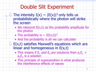
Introduction • One of the great challenges in performing cataract surgery in post-refractive surgery patients is obtaining an accurate refractive outcome, especially in previously myopic patients. • Intraocular lens (IOL) calculation is less reliable in these cases, generally inducing hyperopic errors after cataract surgery. • The main two sources of biometric error are the IOL formulas and the inaccuracy of post-refractive surgery central corneal curvature measurements.
Purpose • To test whether adding Orbscan II calculated values into the double-K method improves IOL calculation in post refractive surgery patients.
Methods A prospective study was undergone with 33 eyes previously submitted to refractive surgery. Post refractive surgery corneal curvature (post-K values) were obtained from the average total-mean power of central 2.0 mm in eyes submitted to myopia correction, and from the central 4.0 mm Orbscan II total mean-power in eyes submitted previously to hyperopia surgery(Bausch&Lomb, Germany). Axial length was obtained from the IOL Master (Zeiss, Germany). The average corneal curvature of the general population (43.8D) was used as the pre-K value into the double-K method. Refraction results 30 days after surgery were compared with refraction that would be obtained if we used: 1) post-K values derived from keratometry; 2) post-K values derived from topography; 3) pre-K average values derived from the central 8 mm area by Orbscan II, and 4) anterior chamber depth measures obtained with the IOL Master and Orbscan II.
Results • Mean post-cataract surgery spherical equivalent was -0.38 ± 1.17 D (range, +1.70 D to -2.97 D) in eyes submitted to myopic surgery, and +0.29 ± 1.77 (range, +2.40 to -1.60 D)in those submitted to hyperopic surgeries.
Results Had we used post-K values derived from keratometer or from topography we would have obtained significantly higher post-operative refractive errors, in eyes previously submitted to myopic refractive surgery (p<0.01). In eyes previously submitted to hyperopic correction, however, there was no statistically significant difference in using keratometry, topography or Orbscan II derived measurements as post-K values. Simulation of results showed there would be less standard deviation of refractive results using topography simulated K values as post-K values in these eyes.
Results Refraction using pre-K average values obtained from the central 8 mm area by Orbscan II were similar to those using the average corneal curvature of 43.8D. Anterior chamber depth measured with IOL Master or Orbscan II did not change the final IOL calculation.
Table 1. Patients data * from 2.0 mm central area in eyes previously submitted to myopic refractive surgery, and of 4.0 mmcentral area in previously hyperopic corrected eyes SE = spherical equivalent RK = radial keratotomy M-Lasik = myopic Lasik H-Lasik = hyperopic Lasik H-PRK = hyperopic photorefractive keratotomy
Table 2. Simulation of results IOL = intraocular lens * from 2.0 mm central area in eyes previously submitted to myopic refractive surgery, and of 4.0 mmcentral area in previously hyperopic corrected eyes SE = spherical equivalent
Conclusion • Average total-mean power of central 2.0 mm Orbscan II used as the post-K values into the double-K method provides a precise way of IOL calculation in post myopic refractive surgery patients.
References • Koch DD, Liu JF, Hyde LL, Rock RL, Emery JM. Refractive complications of cataract surgery after radial keratotomy. Am J Ophthalmol 1989; 108:676-82. • Gimbel HV, Sun R. Accuracy and predictability of intraocular lens power calculation after laser in situ keratomileusis. J Cataract Refract Surg 2001; 27:571-6. • Hoffer KJ. Intraocular lens power calculation after previous laser refractive surgery. J Cataract Refract Surg 2009; 35:759-65. • 4. Ho JD, Liou SW, Tsai RJF, et al. Estimation of effective lens position using a rotating Scheimpflug camera. J Cataract Refract Surg 2008, 34:2119-27. • 5. Qazi MA et al. Determining corneal power using Orbscan II videokeratography for intraocular lens calculation after excimer laser surgery for myopia. J Cataract Refract Surg 2007; 33:21-30. • 6. Borasio E, Stevens J and Smith GT. Estimation of true corneal power after keratorefractive surgery in eyes requiring cataract surgery: BESSt formula. J Cataract Refract Surg 2006; 32:2004-14.7. Aramberri J. Intraocular lens power calculation after corneal refractive surgery: double-K method. J Cataract Refract Surg, 2003; 29:2063-8. • 8. Feiz V, Mannis MJ, Garcia-Ferrer F, Kandavel G, Darlington JK, Kim E, Caspar J, Wang W, Wang W. Intraocular lens power calculation after laser in situ keratomileusis for myopia and hyperopia: a standardized approach. Cornea 2001; 20:792-7. • 9. Sónego-Krone S, López-Moreno G, Beaujon-Balbi OV, Arce CG, Schor P, Campos M. A direct method to measure the power of the central cornea after myopic laser in situ keratomileusis. Arch Ophthalmol 2004; 122:159-66. • 10. Maidana EJ et al. Método para obter el K pré-op usando somente o mapa pós-op. XXV Pan American Congress of Ophthalmology, March 18-21, 2005, Santiago, Chile. • 11. Gelender H. Orbscan II-assisted intraocular lens power calculation for cataract surgery following myopic laser in situ keratomileusis. Trans Am Ophthalmol Soc. 2006; 104:402-13. • 12. Arce CG, Soriano ES, Weisenthal RW, et al. Calculation of Intraocular Lens Power Using Orbscan II Quantitative Area Topography After Corneal Refractive Surgery. J Refract Surg 2009. • 13. Savini G, Barboni P, Profazio V. Corneal power measurements with the Pentacam Scheimpflug camera after myopic excimer laser surgery. J Cataract Refract Surg 2008; 34:809–13. • 14. Packer M, Brown LK, Hoffman RS, et al. Intraocular lens power calculation after incisional and thermal keratorefractive surgery. J Cataract Refract Surg 2004, 30:1430-4. • 15. Shammas HJ, Shammas MC, garabet A, Kim JH, Shammas A, LaBree L. Correcting the corneal power measurements for intraocular lens power calculations after myopic laser in situ keratomileusis. Am J Ophthalmol 2003; 136:426-32. • 16. Garg A. Optical biometry with IOL Master. In: Garg A, Hoyos JE and Dementiev D, eds, Mastering techniques of IOL power calculations. Jaypee Brothers, New Dehli, 2005; 51-54. • 17. Cairns G and McGhee CNJ. Orbscan computerized topography: attributes, applications, and limitations. J Cataract Refract Surg 2005, 31:205-20. • 18. Reddy AR, Pande MV, Finn P, El-Gogary H. Comparative estimation of anterior chamber depth by ultrasonography, Orbscan II, and IOL Master. J Cataract Refract Surg 2004, 30:1268-71. • 19. Wang L, Booth MA, Koch DDl. Comparison of intraocular lens power calculation methods in eyes that have undergone LASIK. Ophthalmology 2004, 111:1825-31. • 20. Shammas HJ and Shammas MC. No-history method of intraocular lens power calculation for cataract surgery after myopic laser in situ keratomileusis. J Cataract Refract Surg 2007; 33:31-6. • Haigis W. Intraocular lens calculation after refractive surgery for myopia: Haigis-L formula. J Cataract Refract Surg 2008; 34:1658-63. • Masket S and Masket SE. Simple regression formula for intraocular lens power adjustment in eyes requiring cataract surgery after excimer laser photoablation. J Cataract Refract Surg 2006; 32:430-4