Download
1 / 27
290 likes | 473 Vues
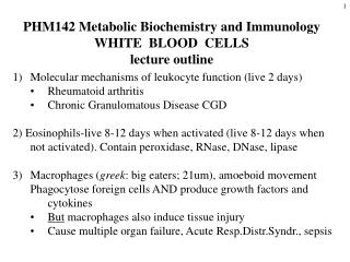
PHM142 Metabolic Biochemistry and Immunology WHITE BLOOD CELLS lecture outline. Molecular mechanisms of leukocyte function (live 2 days) Rheumatoid arthritis Chronic Granulomatous Disease CGD

E N D
PHM142 Metabolic Biochemistry and Immunology WHITE BLOOD CELLS lecture outline • Molecular mechanisms of leukocyte function (live 2 days) • Rheumatoid arthritis • Chronic Granulomatous Disease CGD 2) Eosinophils-live 8-12 days when activated (live 8-12 days when not activated). Contain peroxidase, RNase, DNase, lipase • Macrophages (greek: big eaters; 21um), amoeboid movement Phagocytose foreign cells AND produce growth factors and cytokines • But macrophages also induce tissue injury • Cause multiple organ failure, Acute Resp.Distr.Syndr., sepsis
The Immune System • Two branches: innate/ nonspecific and adaptive/specific • In the innate system: mast cells, neutrophils and macrophages (engulf cytokines inflammation) • Within adaptive, two branches: humoral-mediated (B cells) and cell-mediated (T cells) • Macrophages have a role in both branches
Blood Cells Main function Transport O2 and CO2 Destroy invading bacteria and microbes Phagocytose and kill inavading pathogens Destroy parasites and modulate allergic inflammatory reactions. Release histamine, serotonin, bradykinin, heparin, and cytokines; converts arachidonic acid to prostaglandins & leukotrienes (T & B cells) mediate cytokine release Phagocytose & kill ingested microbes Kill virally infected cells and tumor cells (offer “natural” immunity as well as adaptive) Initiate blood clotting; also release histamine and serotonin (5HT) Type of cell Red blood cells (erythrocytes) White blood cells (leukocytes): Polymorphonuclear/granular leukocytes: Neutrophils (50 60%) Eosinophils (1 4%) Basophils (0.5 2%) Mononuclear leukocytes: Lymphocytes (20 40%) Monocytes (2 9%) ( macrophages in extravascular tissue), Killer cells Megakaryocytes ( platelets)
2 1 Macrophage 3 Thin sections of the major types of white blood cells (leukocytes) found in the circulation, showing the variety of internal structures observed.
Inflammation or infection MPO(myeloperoxidase), HOCl(hypochlorite), SOD (superoxide dismutase). HOCl + red cell GSH = GSH cyclic sulfonamide (biomarker for HOCl formation in vivo that reflects inflammation (oxidant formed by neutrophils)) Living with a killer: The effects of hypochlorite/hypobromite on mammalian cells Chem.Res.Toxicol.21,1011-6 (2008)
Molecular mechanisms of neutrophil function 1 Amer J Med. 109, 33-44 (2000) Neutrophil membrane electron transport CYTOSOL PHAGOSOME MEMBRANE 2 2 PHAGOSOME VACUOLE Fe2+
Activation of leukocyte NADPH oxidase (Nox) showing assembly of the enzyme and fusion of the oxidase-containing vesicle with the phagosomal membrane. Babior, 2000. American Journal of Medicine. 109:33 44.
NADPH OXIDASE DEFICIENCY Chronic Granulomatous Disease (CGD) White Cell bact. 0.1 M Chloride in blood • CGD can kill Bacteria that produce H2O2 and have low catalase activity, e.g., streptococci. • But can’t kill bacteria which have high catalase activity or low H2O2 production, e.g., serratia, nocardia, and aspergillus; fungal, staphylococcus, burkholderia (pneumonia, sepsis).
NADPH oxidase family - makes antibacterial H2O2 • NOX1 – colon > prostate, uterus, breast,macrophage • NOX2 - phagocyte H2O2 for MPO >> hepatocyte, B lymphocyte, cardiomyocytes, endothelial cells • NOX3 inner ear, fetus • NOX4 kidney, bloodvessels,cardiomyocytes,endoth. • NOX5 lymphoid tissue,testis
Rheumatoid Arthritis is an autoimmune inflammatory disease that may affect (skin, blood vessels, heart, lungs, muscles-- but principally attacks joints (proliferative synovitis progressing to cartilage destruction/joint ankylosis. Pathogenesis (unknown etiology):genetic susceptibility; joint damage mediated by leukocytes or exogenous arthritogen (virus, mycobacteria). leukocytes Synovium (synovial fluid) Babior, 2000. American Journal of Medicine. 109:33 44. From Am J Med. (2000). 190:33-44
NSAID therapy for RA IBUPROPHEN COX-1 inhib. 300mg 3 times perday Cardiovascular & GI bleeding risk,inhib platelets, kidney/liver tox. ALEVE (Naproxen) 200mg otc ; lasts 12h • Inhibits inflammation , COX-I and COX-2 inhibitor • Decrease pain, temp, muscle pain, menstrual cramps • Lower stroke risk than ibuprofen; high dose risk GI bleed. VIOXX COX-2 inhibitor for OA,RA 1999 introd. • 2004 (withdrawn due to heart attack, stroke) CELEBREX (celecoxib)1998 COX-2 inhib.400mg • low cardiorenal tox, platelet effects & GI bleeding
Side effects of COX-1 inhibitors • e.g. Aspirin may cause stomach bleeding and risk GI ulcer formation as a result of stomach cell mitochondrial uncoupling and acidosis. • inhibits COX-1 activity thereby increasing tissue unsat. fatty acid levels and causing acidosis. • decreases PGE2 levels that protect stomach membr. • inhibits thromboxane formn. and platelet aggreg. • unsat.fatty acid + PGS(prostaglandin synthase) attacks protective mucous layer
Mechanism of NSAID induced GI toxicity • Stage I NSAIDs decrease intestinal mucosal prostanoids (PGE,TXB2, 6-keto-PGF) • Mitochondrial uncouplers (e.g.indomethacin,DNP) compromised intestinal barrier). Resp.inhibited • Stage 2 mild inflammation + aspirin • Stage 3 histopathology, ulcers and bleeding • Rat model: ulcers, intestinal inflammation or gastric permeability induced by indomethacin or DNP + Cox inhib (aspirin). Aliment Pharmacol. Ther. (2000) 14,639-650
Rheumatoid Arthritis Therapy Repetitive hypoxia in joints, and endothelial cells ATP hydrolysis Hypoxanthine from ATP Reperfusion O2 + endothelial cell xanthine oxidase ALLOPURINOL Monosodium urate crystals in joint Uric acid ALLOPURINOLinhib. ROS endothelial cell nitric oxide Destruction of joint peroxynitrite
Anti-Inflammation therapy • ROS Scavenger Therapy SOD, selenomethionine/Vit E, 5-aminosalicylate, penicillamine:Cu • Macrophage inhibitor therapy: Gold thiomalate or auranofin or zinc or copper salicylate • Prostaglandin Synthetase Inhibitors:-(NSAIDS) COX-1 many cells e.g.aspirin,ibuprofen, but GI bleeding,kidney COX-2 inflammatory cells e.g. VIOXX (withdrawn), CELEBREX but cardiovascular problems. • “Biologics” antibodies that inhibit inflammatory cytokines e.g.TNF-α • Diet: decrease arachidonate intake (meat), increase omega 3 fatty acids (fish) decreases bad prostaglandins, decrease Fe intake References: Semin. Arthritis Rheum. 27: 366-70 (1998). Autoimmunity Reviews 7,1-7(2007) Anti-inflammatory Biologics
PART 2 - EOSINOPHILS: (chemical warfare) Accumulate in: parasitic infections, asthma, rheumatoid diseases, Hodgkin’s lymphoma and allergic or inflammatory diseases • destroy parasitic worms, tumor cells, fungi and bacteria by forming hypobromite H2O2 + Br- + H+ HOBr (hypobromite) + H2O 2) cytokine production e.g. PAF, LTC4 unlike neutrophils (leukocytes) . MPO Biochem J. 358, 233-239 (2001), Journal of Biological Chemistry270 (7) 2905-2913(2000).
Part 3 - Macrophages • White blood cells within tissue, have a role in innate and adaptive immunity • They engulf pathogens and debris via phagocytosis, and move around via amoeboid movement
MACROPHAGES - chemical warfare function 1) Endocytosis and exocytosis via specific receptors for IgG and C3 coated in bacteria 2) H2O2production by NADPH oxidase to kill mycobacteria 3) Arachidonate oxidation to prostaglandin 4) Cytokine production - upon activation by PDGF a) lipopolysaccharide (endotoxin) TNF-a b) immune system activation BCG infection IL-1 c) inflammation or interferon (IFN-g) PAF TGF a and b arginine nitric oxide kill tumor cells 5) endocytosis and delivery to lysosomes (via scavenger receptor) of oxidised LDL (low density lipoprotein) - can result in transformation to foam cell (the basis of the formation of atherosclerotic plaque)
Tumor Necrosis Factor (TNFα) as the primary trigger for inflammatory response Macrophages,monocytes,lymphocytes,keratinocyte • TNFα incr in chronic inflammatory diseases: rheumatism,arthritis,encephalitis,tumors • Rheumatoid arthritis , psoriasis,Crohn’s disease • Proinflammatory > antiinflammatory cytokines • DRUG THERAPY: NSAIDs, GC glucocorticoids, Disease Modifying Antirheumatic Drugs (DMARDs).
Drug induced hepatocyte cytotoxicity caused by activated immune cells releasing cytokines and reactive oxygen species (Kupffer cells , macrophages ,neutrophils) i 1) Toxic doses of drugs or chemicals injure hepatocytes. Injured hepatocytes release factors that attract Kupffer cells to specific regions of the liver. 2) Additional mononuclear phagocytes are also recruited from blood and bone marrow precursors. 3) Once localised in the liver, the macrophages become activated by hepatocyte-derived factors of endothelial cells. 4) Activated macrophages and endothelial cells release cytokines e.g.TNFα & platelet activating factor prime & activate Kupffer cells which release Reactive Oxygen Species and more cytokines. 5) Some chemoattractants and cytokines can attract and activate neutrophils that also contribute to hepatocyte injury.
Macrophages and Tissue Injury Toxicant Target tissues Activated macrophages e.g. Kupffer cells Mediators Gold thiomalate Gadolinium chloride (macrophage inhibitor) Amplification Cytotoxicity Tissue Injury Model for the role of macrophages in tissue injury by generating inflammatory mediators.
Macrophage killing mechanism • Macrophage oxid.LDL which are then endocytosed by the oxid LDL receptor. Present antigens to T cells as foreign substances • Exocytose (via specific receptors) IgC and C3 coated bacteria • H2O2 kills mycobacteria and form cytokines PDGF,PAF,TNFα & β, IL1 • Unsaturated fatty oxidation to aldehydes causes foam cell formation & plaque formation from macrophages.
Inflammatory mediators implicated in toxicity Toxicant Mediator Lung Liver 1) Reactive oxygen intermediates Ozone Endotoxin (H2O2, .OH) Asbestos Acetaminophen Amiodarone Corynebacterium parvum Reactive nitrogen intermediates Bleomycin (peroxynitrite) Carbon tetrachloride 1,2-dichlorobenzene Phenobarbital Endotoxin 2) Hydrolytic enzymes Endotoxin (collagenase, elastase) Silica 3) Lipids Hyperoxia (leukotrienes, prostaglandins, thromboxanes) 4) IL-1 Cigarette smoke 5) TNF-a (mitochondrial toxin, Cadmium chloride Alcohol reactive oxygen species) Toxicology 160, 111 8 (2001). Ann. Rev. Pharmacol. Toxicol. 35, 655 (1995).
4. Biologics: anti-cytokine antibodies • TNFa blockers 1)Etanercept for RA • 2) Infliximab (remicade)Crohns disease, • drug antigen?, cancer a chimeric human Fab. 3) Adalimumab (Humira) human monoclonal for macular degen. 4)Stelera for psoriasis, 5 shots p.a.$50K • Rituximab kills ANCA-vasculitis NEJM363,221-232(2010) & CD20 B cell,NH lymphoma,RA, Wegeners ? • Abatacept,fusion protein inhib.T cell costim.RA • Avastin (bevacizumab) inhib.VEGF-A (vascular endoth.growth factor,eye mac.degen, breastcancer).Withdrawn.