DNA Structure and Replication
450 likes | 535 Vues
Explore the journey of DNA discovery and replication through critical experiments like Griffith's & Hershey-Chase. Understand the structure & significance of DNA in genetic heredity.

DNA Structure and Replication
E N D
Presentation Transcript
DNA Structure and Replication Honors Biology
What you will learn… 1. Discovery of Genetic Material 2. DNA Structure 3. The Race to Solve the Puzzle of DNA Structure 4.DNA Replication
1. Disovery of Genetic Material: a. DNA or Protein? • Biologists knew that genes are located on chromosomes (made of DNA and protein) • DNA and protein were the candidates for the genetic material • Until the 1940s, the case for proteins seemed stronger because proteins appeared to be more structurally complex and functionally specific. • Biologists finally established the role of DNA in heredity through studies involving bacteria and the viruses that infect them.
1. Discovery of Genetic Materialb. Griffith Experiment • 1928; British medical officer • Griffith was studying two strains of a bacterium: • a pathogenic (disease-causing) strain that cause pneumonia • a harmless strain.
1. Discovery of Genetic Materialb. Griffith Experiment • Found that when he mixed a dead version of the pathogenic bacteria and harmless bacteria, some living bacterial cells were converted to the disease-causing form. • Furthermore, all of the descendants of the transformed bacteria inherited the newly acquired ability to cause disease.
1. Discovery of Genetic Materialb. Griffith Experiment Clearly, some chemical component of the dead bacteria could act as a “transforming factor” that brought about a heritable change.
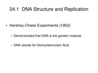
1. Discovery of Genetic Materialc. Hershey Chase Experiment 1952; American biologists Experiments showed that DNA is the genetic material of a virus (bacteriophage or phage, for short) called T2, which infects E.coli T2 consists solely of DNA and protein; DNA-containing head and a hollow tail with six jointed fibers extending from it.
1. Discovery of Genetic Materialc. Hershey Chase Experiment • T2 infects bacteria by attaching to the surface with its fibers and injecting its hereditary material. • Raised the question: Is it DNA being passed on or protein? • To answer protein or DNA question, they devised an experiment to determine what kinds of molecules the phage transferred to E.coli during infection
1. Discovery of Genetic Materialc. Hershey Chase Experiment • Used a few relatively simple tools: • Chemicals containing radioactive isotopes • To label the DNA and protein in T2 • Used radioactive sulfur and phosphorous • Sulfur is in proteins. Phosphorous is in DNA. • A radioactivity detector • A kitchen blender • And a centrifuge (device that spins test tubes to separate particles of different weights.
1. Discovery of Genetic Materialc. Hershey Chase Experiment • The Experiment • First they grew T2 with E.Coli with radioactive sulfur. • Then, they grew a separate batch of phages in a solution containing radioactive phosphorous. • They allowed the two batches of T2 to infect separate samples of nonradioactive bacteria • Shortly after the onset of infection, they agitated the cultures in a blended to shake loose any parts of the phages that remained outside the bacterial cells. • They then spun the mixtures in a centrifuge. The cells were deposited as a pellet at the bottom of the centrifuge tubes, but phages and parts of phages being lighter, remained suspended in the liquid. • The researchers then measured the radioactivity in the pellet and the liquid.
1. Discovery of Genetic Materialc. Hershey Chase Experiment • The Results • Found that when the bacteria has been infected with T2 phages containing labeled protein, the radioactivity ended up in the liquid but not bacteria. • Result suggested that the phage protein did not enter the cells. • But when the bacteria had been infected with phages whose DNA was tagged, then most of the radioactivity was in the pellet, made up of bacteria. • When these bacteria were returned to liquid growth medium, they soon died and lysed and released new phages that contained radioactive phosphorous in their DNA but no radioactive sulfur.
1. Discovery of Genetic Materialc. Hershey Chase Experiment • The Conclusions • They concluded that T2 injects its DNA into the host cell, leaving virtually all its protein outside. • They demonstrated that it is the injected DNA molecules that cause cells to produce additional phage DNA and proteins, making new complete phages. • This indicated that DNA contained the instructions for making proteins • ***THESE RESULTS CONVINCED MOST SCIENTISTS THAT DNA IS THE HEREDITARY MATERIAL!!!***
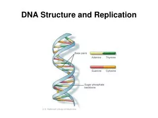
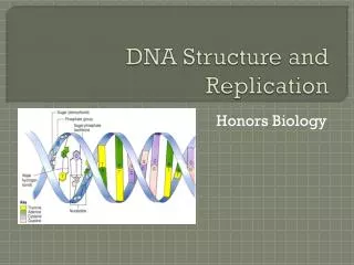
2. DNA Structurea. General • Deoxyribonucleic Acid (DNA) • Monomers made up of nucleotides: • Nucleotides consist of: • A five carbon sugar, deoxyribose • Phosphate group • Nitrogenous base (Adenine, Guanine, Cytosine, Thymine) • Double helix consists of: • Sugar-phosphate backbone held by covalent bonds • Nitrogen bases are hydrogen bonded together; A pairs with T and C pairs with G
2. DNA Structureb. Nucleotide structure • Nucleotides consist of: • A five carbon sugar, deoxyribose • Four in it’s ring, one extending above the ring • Missing one oxygen when compared to ribose • Phosphate group • Is the source of the “acid” in nucleic acid • Nitrogenous base (Adenine, Guanine, Cytosine, Thymine) • A ring consisting of nitrogen and carbon atoms with various functional groups attached • Double ring= purines (A and G) • Single ring= pyrimidines (T and C)
2. DNA Structurec. 5’ end and 3’ end DNA’s sugar-phosphate backbones run in opposite directions. Each strands has a 3’ end and a 5’ end. The primed number is referring to the carbon atoms of the nucleotide sugars. At one end of each DNA strand, the sugar’s 3’ carbon atom is attached to an –OH group, at the other end, the sugar’s 5’ carbon has a phosphate group.
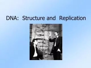
3. The Race to solve the puzzle of DNA structure A few scientists working on the puzzle trying to determine the 3-D structure: Franklin, Watson and Crick Rosalind Franklin observed an X-ray crystallography image of the basic shape of DNA
3. The Race to solve the puzzle of DNA structure • Watson saw this image, and with just a glance deduced the basic shape of DNA to be a helix with a uniform diameter of 2 nm, with its nitrogenous bases stacked about one-third of a nanometer apart. • The diameter of the helix suggested it was made up of two polynucleotide strands=> DOUBLE HELIX!
3. The Race to solve the puzzle of DNA structure • Watson and Crick began trying to construct a double helix that would conform both Franklin’s data and what was currently known about the chemistry of DNA. • Franklin had concluded that the sugar-phosphate backbones must be on the outside of the double-helix, forcing the nitrogenous bases to swivel to the interior of the molecule.
3. The Race to solve the puzzle of DNA structure Watson and Crick found that Adenine always paired with Thymine, and Guanine and Cytosine, to ensure a uniform diameter. Complementary base pairing was explained both by the physical attributes and chemical bonding of DNA, along with data obtained by Chargaff
3. The Race to solve the puzzle of DNA structure • Chargaff’s rules: A always pairs with T and G always pairs with C. • Only apply to base pairing, not the sequence of nucleotides • The sequence of bases can vary in countless ways, and each gene has a unique order of nucleotides, or base sequence
3. The Race to solve the puzzle of DNA structure 1962, Watson and Crick received the Nobel Prize for their work (Franklin would have received it as well, but she died from cancer in 1958; Nobel Prizes are never awarded to the deceased)
4. DNA Replication “It has not escaped our notice that the specific pairing we have postulated immediately suggests a possible copying mechanism for the genetic material” ~Watson and Crick
4. DNA Replication • Logic behinds Watson-Crick’s proposal for how DNA is copied • Can be seen by covering one of the strands in the parental DNA molecule with a piece of paper: you can determine the bases of the covered strand by applying the base-pairing rules: A pair with T, and G pairs with C. • Watson and Crick predicted that a cell applies the same rules when copying its genes.
4. DNA Replicationa. Overview • Figure 10.4A Template model for DNA replication • First, the two strands of parental DNA separate, and each becomes a template for the assembly of a complementary strand from a supply of free nucleotides. • The nucleotides line up one at a time along the template strand in accordance with the base-pairing rules • Enzymes link the nucleotides to form the new DNA strands. • Completed new molecules, identical to the parental molecule, are known as daughter DNA.
4. DNA Replicationa. Overview • Semi-conservative model • Watson and Crick’s model predicts that when a double helix replicates, each of the two daughter molecules will have one old strand, which was part of the parental molecule, and one new made strand. • Known as semi-conservative model because half of the parental molecule is maintained (conserved) in each daughter molecule. • Confirmed by experiments performed in the 1950s.
4. DNA Replicationa. Overview The opposite orientation of the strands is important in DNA replication. DNA polymerases link DNA nucleotides to a growing daughter strand, only to the 3’ end of the strand, never to the 5’ end. Thus, a daughter DNA strand can only grow in the 5’3’ direction.
4. DNA Replicationa. Overview • We will be looking at three steps in DNA Repication: • 1. Initiation • 2. Elongation • 3. Termination
4. DNA Replication b. Intiation • Initiation: • DNA replication begins at specific sites on the double helix referred to as origins of replication. • An enzyme called helicase binds to DNA and separates the strands. • Uses energy from ATP • Single-strand binding protein (SSB) binds to each strand to prevent reannealing
4. DNA Replicationb. Initiation • Initiation (continued) • Replication proceeds in both directions , creating replication “bubbles” • DNA has many origins of replication that can start simultaneously, for time efficiency. • Thousands of bubbles can be present, and eventually all the bubbles merge, yielding two completed daughter DNA molecules.
4. DNA Replicationc. Elongation • Elongation: • Replication fork forms: • Partial opening of a DNA helix to form two single strands that has a fork appearance. • Primers , made of short stetches of RNA, are required byDNA polymerases during replication • synthesized by an enzyme called RNA primase.
4. DNA Replicationc. Elongation • Elongation (continued): • DNA polymerase removes primers and adds DNA nucleotides. • We will be looking at two types of DNA Polymerase: DNA polymerase III and DNA polymerase I
4. DNA Replicationc. Elongation • Elongation (continued): • one of the daughter strands can be synthesized in one continuous fashion from an initial primer, working toward the forking point of the parental DNA. leading strand • Only a single priming event is required, and then the strand can be extended indefinitely by DNA polymerase III.
4. DNA Replicationc. Elongation • Elongation (continued): • The other daughter strand polymerase molecules must work outward from the forking point, is synthesized in short pieces as the fork opens up in a discontinous fashion involving multiple priming events. lagging strand
4. DNA Replicationc. Elongation • Elongation (continued): • Lagging strand (continued) • DNA Polymerase III adds nucleotides in 5’3’ direction. Primersget removed via and replaced by DNA via DNA polymerase I • Fragments formed are called Okazaki fragments • DNA ligase links (ligates) the pieces together into a single DNA strand.
4. DNA Replicationd. Termination • Termination: • At the completion DNA replication, you end up with two identical strands of DNA. • Each DNA molecule contains one original parent strand, and one new daughter strand. Semi conservative replication
4. DNA Replication (animation) http://www.wiley.com/college/pratt/0471393878/student/animations/dna_replication/index.html
DNA Replicatione. key enzymes • Key enzymes: • Helicase: unwinds the double helix • Primase: synthesizes RNA primers • SSB: stabilizes single-stranded regions; prevents reannealing • DNA polymerase III- synthesized DNA • DNA polymerase I- erases primer and fills gaps • DNA ligase- joins the ends of DNA segments; DNA repair
4. DNA Replicatinf. Checking mechanisms • DNA polymerase carry out a proofreading step that quickly removes nucleotides that have base-paired incorrectly • DNA polymerases and DNA ligase are also involved in repairing damaged DNA by harmful radiation or toxic chemicals • Process is not only fast but also amazingly accurate • Typically, only about one DNA nucleotide per billion is incorrectly paired • Ensure that all somatic cells in a multicellular organism carry the same genetic information.