Download
1 / 97
990 likes | 1.11k Vues
Explore the various techniques for determining and purifying organic compounds, including distillation, crystallization, and chromatography. Learn how these methods work and their applications in organic chemistry research.

E N D
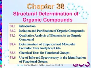
Determination of organic structures ? Product
Product purification Distillation Crystallization • Chromatography • Thin-layer chromatography (TLC) • Column (flash) chromatography • Gas or liquid chromatography
Distillation Distillation works by exploiting the different boiling points of liquids. The more volatile liquid (lower b.p.) will simply evaporate first and the vapor will pass into a condenser where it reverts into liquid. Further heating will force the less volatile liquids (higher b.p.) to evaporate and distill at higher temperature.
Crystallization Crystallization is amethod for purifying solids. Crystallization works by taking advantage of the different solubility of compounds, and allows impurities to be removed from crude solids. First, you add a small amount of solvent to the crude compound in a flask, and heat toboiling until the solid has dissolved. Then, you cool the solution, allowing crystals to precipitate, while impurities stay dissolved in the solvent. Then you separate crystals by vacuum filtration, and discard the waste solution.
Chromatography Methods of compounds mixture separation based on the different distribution of its components between two phases: stationary phaseand mobile phase The mobile phase can be a liquid or a gas and the stationary phaseis usually a solid or nonvolatile liquid. Combinations of these two components result in different chromatographic techniques.
Column chromatography Solvent – mobile phase Stationary phase (SiO2 or Al2O3) Glass wool The glass tube is filled with adsorbent material, and solution containing the mixture to be separated is allowed to drip through the column
Thin Layer Chromatography Here the mobile phase is a liquid Stationary phase is a thin layer of a solid on a flat surface (glass plate). Substances that are weakly adsorbed onto the solid or are more soluble in the liquid move faster. And so move further up the plate by the time that the process has been stopped by taking the plate out of the liquid.
TLC – Thin Layer Chromatography Spots visualization is attained by spraying the plate with reagent forming coloured compounds with analyzed substances Method used for determining the purity of materials and also for preliminary identification purposes Adsorbent is supported as a thin coating on a flat surface (glass; aluminum plate)
b a Distance moved by the substance [a] Distance moved by the solvent [b] TLC – RF (distribution coefficient) Rf=
Controlling the reaction progress by TLC Reaction finished by-products present 1st line – substrate standard 2nd line – reaction mixture 3rd line – product standard Reaction finished - OK
H RESET Air Hydrogen Gas Carrier Gas Chromatography Filters/Traps Data system Regulators Syringe/Sampler Inlets Detectors • gas system • inlet • column • detector • data system Column
Gas Chromatography Capillary column Stationary solid phase column
Gas Chromatography Applications QUALITATIVE AND QUANTITATIVE ANALYSIS OF COMPLEX MIXTURES
Gas Chromatography Applications TENTATIVE IDENTIFICATION OF UNKNOWN COMPOUNDS
Gas chromatographyadvantages • 1. Very good separation • 2. Time of analysis is short • 3. Small sample is needed - l or g • 4. Good detection systems • Qualitative and quantitative analysis
Gas Chromatography drawbacks 1. Sample has to be volatilized at 250°C without decomposition 2. Nonvolatile compounds can be transformed into stable derivatives but it is: time consuming side reaction can accompany loss of sample
High Performance Liquid Chromatography Once called High Pressure Liquid Chromatography
What is HPLC? • The widely used analytical separations technique • Utilizes a liquid mobile phase to separate components of mixture • Uses high pressure to push solvent through the stationary phase in the column
HPLC is…. • sensitive method • ready adaptable to accurate quantitative analysis • suitable for separating nonvolatile species orthermally unstable ones • ideally suited for separation and identification of biomolecules - amino acids, proteins, nucleic acids, carbohydrates, as well as hydrocarbons, pharmaceuticals, pesticides, pigments, antibiotics, steroids, and a variety of other substances
Parts of HPLC apparatus HPLC column HPLC injector
Advantages of HPLC • High resolution and short time of analysis • HPLC columns can be reused without repacking or regeneration • Greater reproducibility due to close control of the parameters affecting the efficiency of separation • Easy automation of instrument operation and data analysis • Adaptability to large-scale, preparative procedures
What is spectroscopy?? ? ? Spectroscopy is the use of the absorption, emission, or scattering of electromagnetic radiation by matter to qualitatively or quantitatively study the matter or to study physical processes. The matter can be atoms, molecules, atomic or molecular ions, or solids. The interaction of radiation with matter can cause redirection of the radiation and/or transitions between the energy levels of the atoms or molecules. Absorption: A transition from a lower level to a higher level with transfer of energy from the radiation field to an absorber, atom, molecule, or solid. Emission: A transition from a higher level to a lower level with transfer of energy from the emitter to the radiation field. If no radiation is emitted, the transition from higher to lower energy levels is called nonradiative decay. Scattering: Redirection of light due to its interaction with matter.
Spectroscopic methods used for structure determination Mass spectrometry (MS) What is molecular formula? Infrared spectroscopy (IR) What functional groups are present? Ultraviolet spectroscopy (UV-VIS) Is a conjugated π-electron system present? Nuclear magnetic resonance spectroscopy (NMR) What carbon-hydrogen (1H-NMR); carbon-carbon (13C-NMR) framework is present? X-ray spectroscopy What are bond lenghts and bond angles? Usually, no one single spectroscopic method is reliable by itself for determining the structure of an unknown. A combination of two or more methods is almost always required
Mass spectrometry • A mass spectometer has three basic components: • Something to volatilize and ionize the molecule into a beam of charged particles • Something to focus the beam of particles of the same mass • Something to detect the particles • Two methods are used to convert molecules into ions (usually positive – cations) • electron impact • chemical ionization • Other methods for ionization • Plasma and glow discharge • Electrospray ionization • Fast atom bombardment (FAB) • Field ionization – uses high energy field • Laser ionization • Matrix assisted lasser desorption ionization • Plasma desorption ionization • Resonance ionization • Secondary ionization • Thermal ionization
In electron impact MS the molecule is bombarded with high energy electrons that knock a weakly bound electron out of molecule. Losing a single electron leaves behind a radical cation: an unpaired electron and a positive charge. Some of these rather unstable radical cations will survive the focusing operation and get to the detector. Molecule first forms the radical cation M+, which then breaks up (fragments) to give radical X* and a cation Y+. Only charged particles can be focused by the magnetic field and do the detector records only ion M+, and positively charged fragments Y+. Uncharged radicals are not recorded.
A mass spectrum will usually be presented as a vertical bar graph, in which each bar represents an ion having a specific mass-to-charge ratio (m/z) and the length of the bar indicates the relative abundance of the ion. The most intense ion is assigned an abundance of 100%, and it is referred to as the base peak. Most of the ions formed in a mass spectrometer have a single charge, so the m/z value is equivalent to mass itself. Modern mass spectrometers easily distinguish (resolve) ions differing by only a single atomic mass unit (amu), and thus provide completely accurate values for the molecular mass of a compound. The highest-mass ion in a spectrum is normally considered to be the molecular ion, and lower-mass ions are fragments from the molecular ion, assuming the sample is a single pure compound. m/z peak intensity n-hexane 15.0 1.3 26.0 1.7 27.0 22.7 28.0 4.7 29.0 42.5 39.0 14.8 40.0 3.1 41.0 72.5 42.0 42.4 43.0 80.9 44.0 2.8 55.0 12.1 56.0 70.5 57.0 100.0 58.0 4.7 69.0 8.5 70.0 2.6 71.0 11.0 84.0 3.2 86.0 10.0
High Resolution Mass Spectroscopy By designing mass spectrometers that can measure m/z values accurately to four decimal places, it is possible to distinguish different molecular formulasof the same nominal mass.
Hexane shows the same fragmentation pattern as other unbranched alkanes. Thus, alkyl carbocations at m/z=15, 29, 43 and 57 amu provide the dominant peaks in the spectrum. The m/z=57 butyl cation (M-29) is the base peak, and the m/z=43 and 29 ions are also abundant Chain branching clearly influences the fragmentation of this isomeric hexane. The molecular ion at m/z=86 is weaker than that for hexane itself and the M-15 ion at m/z=71 is stronger. The m/z=57 ion is almost absent (try to find a simple cleavage that gives a butyl group). An isopropyl cation (m/z=43) is very strong, and the corresponding propene radical-cation at m/z=42 (colored orange), produced by loss of propane, gives the base peak. By having the six carbons of hexane closed to a ring, the fragmentation is profoundly changed. To begin with, the molecular ion at m/z=84 is much stronger than the corresponding ions in the previous acyclic compounds. The base peak at m/z=56 is produced by loss of ethene, so it is an odd-electron ion (colored orange). The alkenyl cations at m/z=41 & 27 are stronger than the corresponding alkyl cations (m/z=43 & 29). The loss of methyl (m/z=69), and a corresponding small m/z=15 ion obviously require some hydrogen rearrangements.
GC+MSLC+MS+computer libraries of spectraenable qualitative and quantitative analysis of complex mixtures even when very small samples are accessible
Infrared spectroscopy (IR) What functional groups are present?
INFRARED SPECTROSCOPY IR range λ=2.5x10⁻⁴ cm - 2.5x10⁻³ cm /// 2.5 - 25μm ν= 4000 - 400 [cm-1] (wavewnumber) FUNCTIONAL GROUP FINDER
Infrared spectroscopy detects the stretching and bending of bonds Photon energies associated with this part of the infrared (from 1 to 15 kcal/mole) induce vibrational excitation of covalently bonded atoms and groups. The covalent bonds in molecules are not rigid sticks or rods, such as found in molecular model kits, but are more like stiff springs that can be stretched and bent
symmetric stretching antisymmetric stretching In-plane bending Out-of-plane bending The amount of energy needed for stretching and bending of bondscorresponds infrared weavelenghts IR spectra are simple absorption spectra Wavelength energy corresponding to a specific wavelength is absorbed, the intensity of the radiation reaching a detector decreases, and this is recorded in the spectrum.
Regions of IR spectra 600-1500 cm-1 fingerprint region 1500- 2000 cm-1 streching of C=C, C=O, C=N bonds 2000-2500 cm-1 streching of C≡C, C≡N bonds 2500-4000 cm-1 streching of C-H, N-H, O-H bonds
cyclohexane 2850-3000 cm-1 C-H stretching 1350-1470 cm-1 C-H bending cyclohexene 1630-1680 cm-1 C=C stretching 3020-3100 cm-1 C=C-H stretching 2850-3000 cm-1 C-H stretching 1350-1470 cm-1 C-H bending benzene 3020-3100 cm-1 C=C-H stretching 1500 cm-1 C=C in ring stretching 690-900 cm-1 C-H bending