Circulatory disorders & Shock
Circulatory disorders & Shock. Jan Laco, M.D., Ph.D. Summary. 1. Edema 2. Hyperemia and Congestion 3. Hemorrhage 4. Thrombosis 5. Embolism 6. Infarction. 1. Edema. = fluid in interstitium cavities – hydrothorax, hydropericardium, ascites anasarca = severe generalized edema

Circulatory disorders & Shock
E N D
Presentation Transcript
Circulatory disorders & Shock Jan Laco, M.D., Ph.D.
Summary • 1. Edema • 2. Hyperemia and Congestion • 3. Hemorrhage • 4. Thrombosis • 5. Embolism • 6. Infarction
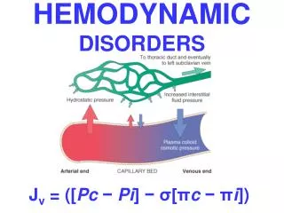
1. Edema = fluid in interstitium • cavities – hydrothorax, hydropericardium, ascites • anasarca = severe generalized edema • 3 major factors: • hydrostatic pressure • plasma colloid osmotic pressure • lymphatic obstruction • inflammation
1. Edema • 1. hydrostatic pressure • impaired venous return • congestive heart failure • constrictive pericarditis • liver cirrhosis – ascites • venous obstruction or compression • thrombosis • external pressure
1. Edema • 2. plasma colloid osmotic pressure • loss or reduced albumin synthesis • nephrotic syndrome • protein-losing gastroenteropathy • liver cirrhosis • malnutrition
1. Edema • 3. lymphatic obstruction • lymphedema • inflammatory – filariasis elephantiasis • neoplastic – breast carcinoma • postsurgical (LN resection) + postirradiation
1. Edema • subcutaneous tissue (pitting edema) + cavities • generalized x local prominent • right heart failure – lower limbs • left heart failure - pulmonary edema • nephrotic syndrome – periorbital edema (eyelids) • brain edema – localized x generalized • gyri flattening + sulci narrowing herniation
2. Hyperemia and Congestion = blood volume in particular tissue • 2a. hyperemia – active (arteriolar dilation) • red • striated muscle exercise • 2b. congestion – passive (impaired venous return) • systemic x local • blue-red color (cyanosis) • accumulation of deoxygenated Hb • chronic chronic hypoxia regressive changes + small hemorrhages siderophages
2. Hyperemia and Congestion • pulmonary congestion • acute • blood fulfilled septal capillaries • septal + alveolar edema + small hemorrhages • chronic • septa thickening fibrosis • alveoli - siderophages (heart failure cells)
2. Hyperemia and Congestion • liver congestion • acute • blood fulfilled central vein + sinusoids • chronic – „nutmeg“ – red-brown + fatty collor • centrolobular necrosis + hemorrhage • periportal fatty change • cardiac fibrosis • bowel congestion • hemorrhagic necrosis
3. Hemorrhage = extravasation of blood from blood vessels • external (+ hollow organ) • within tissue – hematoma • hemorrhagic diatheses – insignificant injury • vasculopathies • trombocytopenia + -patia • coagulopathy
3. Hemorrhage • 1. Petechiae (1-2 mm) - skin + mucosa • intravascular pressure, platelets • 2. Purpuras (3-5 mm) • trauma, vasculitis, vascular fragility • 3. Ecchymosis (1-2 cm) = hematomas (bruises) • – RBC phagocytosis by macrophages • - Hb (red-blue) bilirubin (blue-green) hemosiderin (golden-brown) • 4. Cavities • hemothorax, hemopericardium, hemoperitoneum • hemarthros
3. Hemorrhage • arterial + venous + capillary • H. per rhexin – injury - brain • H. per diabrosin – erosion – peptic ulcer • H. per diapedesin – transmigration of RBC (no damage of capillaries) – toxic injury + stasis
3. Hemorrhage - sequelae • 1. loss volume • > 20% hemorrhagic shock • 2. loss rate • acute hemorrhagic shock • chronic (peptic ulcer, menstrual bleeding) • iron deficiency anemia • 3. site • subcutaneous x brain
Disseminated Intravascular Coagulation (DIC) • basis: widespread activation of thrombin • Mi: fibrin thrombi in microcirculation • 1. stage • multiple fibrin thrombi in microcirculation consumption of PLT + coagulation proteins • 2. stage • fibrinolytic system activation serious bleeding
DIC - causes • 1. obstetric complications • abruption placentae retroplacental hematoma • amniotic fluid embolism • septic abortion • 2. infections • sepsis (Gram +, Gram- bacteria) • meningococcemia • 3. neoplasms • carcinoma of pancreas, prostate, lung, leukemia • 4. tissue injury • burns
4. Thrombosis = intravital intravascular blood clotting • Virchow triad • 1. endothelial injury • physical – hypertension, turbulence • chemical – hypercholesterolemia, smoking, vasculitis • 2. alteration of blood flow • stasis – immobilization, cardiac chamber dilation • 3. hypercoagulability • primary (genetic) x secondary • factor V mutation (Leiden) x neoplasms, drugs,
4. Thrombosis • Grossly: mural x occlusive • Mi: RBC + WBC + PLT + fibrin lines of Zahn - lamination • Sites • arteries + veins + capillaries • cardiac chambers + valve cusps
4. Thrombosis • 1. Arterial thrombi • occlusive • coronary + cerebral + femoral aa. • upon AS plaque + bifurcation • G: gray-white, friable • Mi: PLT + fibrin, RBC + WBC
4. Thrombosis • 2. venous thrombi (phlebothrombosis) • occlusive • deep veins of LL + pelvic plexus • G: firm, red, attached to wall • Mi: RBC + fibrin • !!! asymptomatic (50%) !!! • X postmortal clots (not attached to wall, gelatinous red centre + fat supernatant)
4. Thrombosis • 3. cardiac chambers • upon infarction + dilated cardiomyopathy • mural • 4. valve cusps • infective endocarditis (vegetations) • non-bacterial thrombotic endocarditis (sterile) • Libman-Sacks endocarditis – systemic LE
4. Thrombosis – fate of thrombus • 1. propagation • 2. embolization • 3. dissolution • fibrinolysis (recent thr.) • 4. organization • endothelial cells, smooth muscle cells, fibroblasts, capillaries • 5. recanalization • new small lumina
5. Embolism = detached i.v. solid, liguid or gaseous mass carried by blood to distant site from point of origin • 1. thrombembolism (99%) • pulmonary x systemic infarction • 2. cellular - amniotic fluid, tumor cells • 3. subcellular - AS debries, BM bits • 4. fat • 5. air • 6. foreign bodies - catheter
Pulmonary thrombembolism • source - deep veins of LL + pelvic plexus • v. cava inf. right heart a. pulmonalis • paradoxical embolism - IA or IV defect systemic emboli • + left heart failure pulmonary infarction • large - sudden death (acute right heart failure) • bifurcation – saddle embolus • 60% pulmonary circulation obtructed • small (60-80%) - pulmonary hypertension • branching arterioles fibrinolysis bridging web
Systemic thrombembolism • source: intracardial thrombi (80%) + AS • infarctions • LL (75%) + brain (10%) • bowel + kidney + spleen
Fat embolism • source: fractures of bones with fatty BM + soft tissue trauma + burns • 1. stage (after 1-3 days) • veins lungs pulmonary insufficiency • 2. Stage • lungs systemic circulation neurologic symtoms + thrombocytopenia • 10% fatal • Mi: fat droplets in lung, brain, kidney capillaries
Air embolism • 1. systemic veins lungs • obstetric procedures, goiter operation, chest wall injury • 2. pulmonary veins systemic circulation • cardiosurgery • 100mL of air symptoms (chokes) • air bubbles – physical vessel obstruction • Decompression sickness • deep sea divers (nitrogen) • chronic form – caisson disease – bone necrosis
Amniotic fluid embolism • source: abruptio placentae retroplacental hematoma • a.f. infusion into maternal circulation uterine veins lungs • dyspnea, cyanosis, hypotensive shock, seizures, coma + lung edema + DIC • Mi: pulmonary capillaries (mother) - squamous cells + lanugo hair + fat • + DAD
7. Infarction = ischemic necrosis due to occlusion of arterial supply or venous drainage • causes: • thrombotic or embolic events (99%) • vasospasm, hemorrhage in AS plaque • external compression (tumor) • twisting (testicular + ovarian torsion, bowel volvulus)
7. Infarction - determinants • 1. nature of blood supply • dual – lung + liver • end-arterial – kidney + spleen • 2. rate of occlusion • acute – infarction • chronic – collateral circulation, interart. anastomoses • 3. vulnerability to hypoxia • neurons – 3-4 min • cardiomyocytes - 20-30 min • fibroblasts - hours • 4. oxygen blood content – heart failure, anemia
7. Infarction- morphology • 1. red infarcts • venous occlusion • loose tissue (lung) – blood collection • dual circulation – lung + bowel • previously congested organs • reperfusion (angioplasty, drug-induced thrombolysis) • 2. white infarcts • arterial occlusion • solid organs – heart (yellow), spleen, kidney
7. Infarction- morphology • wedge shape • apex to occluded artery • base to organ periphery + fibrinous exsudate (pleuritis, pericarditis epistenocardiaca) • onset – poorly defined, hemorrhagic • in time – sharper margins + hyperemic rim
7. Infarction- morphology • ischemic coagulative necrosis – 3 zones • 1. total necrosis - centre • loss of nuclei, eosinophilia of cytoplasm, architecture is preserved • 2. partial necrosis • some cells survive • inflammation (neutrophils) – 1-2 day degradation of dead tissue • 3. hyperemic rim
7. Infarction- morphology • healing • granulation tissue (5-7 day) fibrous scar (6-8 weeks) • !!! brain – liquefactive necrosis pseudocyst !
7. Infarction • septic infarctions • source • infective endocarditis (vegetations) • suppurative thrombophlebitis • infarction abscess granulation tissue scar
Shock = systemic hypoperfusion due to reduction of cardiac output / effective blood volume circulation • hypotension cellular hypoxia • features – hypotension, tachycardia, tachypnea, cool cyanotic skin (x septic s. – warm) • initial threat + shock manifestations in organs • prognosis • origin + duration
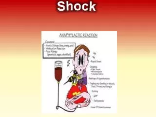
Shock • 1. cardiogenic – failure of myocardial pump • myocardial infarction, arrhythmias • pulmonary embolism • 2. hypovolemic - inadequate blood/plasma volume • hemorrhage • fluid loss (vomiting, diarrhoea, burns, trauma) • 3. septic – vasodilation + endothelial injury • Gram+, Gram- bacteria • 4. neurogenic - loss of vascular tone • spinal cord injury • 5. anaphylactic – IgE–mediated hypersensitivity
Shock - stages • progressive disorder multiorgan failure death • 1. nonprogressive • compensatory mechanism (neurohumoral) activation • centralization of blood circulation • 2. progressive • tissue hypoperfussion – metabolic dysbalancies • 3. irreversible • incurred cellular damage + tissue injury
Shock - morphology • brain - ischemic encephalopathy • tiny ischemic infarctions (border zones) • heart • subendocardial hemorrhage + necroses, contr. bands • kidney - acute tubular necrosis (shock kidney) • pale, edematous • tubular epithelium necroses casts • lung – diffuse alveolar damage (shock lung) • heavy, wet • congestion + edema + hyaline membranes
Shock - morphology • adrenal gland • lipid depletion • GIT – hemorrhagic enteropathy • mucosal hemorrhages + necroses • liver • fatty change, central necrosis