Download
1 / 44
440 likes | 468 Vues
Understand the composition, adhesion, and transport processes in cellular membranes. Learn about endocytosis, exocytosis, and dynamic membrane properties. Figures and tables offer visual aids for better comprehension.

E N D
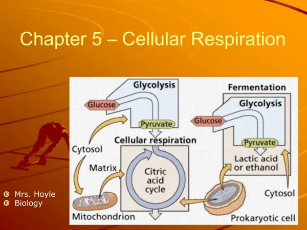
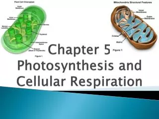
Chapter 5: Cellular Membranes Membrane Composition and Structure Cell Adhesion Passive Processes of Membrane Transport Active Transport
Chapter 5: Cellular Membranes Endocytosis and Exocytosis Membranes Are Not Simply Barriers Membranes Are Dynamic
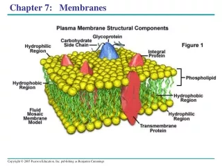
Membrane Composition and Structure • Biological membranes consist of lipids, proteins, and carbohydrates. • The fluid mosaic model describes a phospholipid bilayer in which membrane proteins move laterally within the membrane. Review Figures 5.1, 5.2 4
figure 05-01.jpg 5.1 Figure 5.1
figure 05-02.jpg 5.2 Figure 5.2
Membrane Composition and Structure • Integral membrane proteins are partially inserted into the phospholipid bilayer. • Peripheral proteins attach to its surface by ionic bonds. Review Figure 5.1 7
Membrane Composition and Structure • The two surfaces of a membrane may have different properties due to different phospholipid compositions, exposed integral membrane proteins, and peripheral membrane proteins. • Defined regions of a plasma membrane may have different membrane proteins. Review Figures 5.1, 5.2 8
Membrane Composition and Structure • Carbohydrates attached to proteins or phospholipids project from the external surface of the plasma membrane and function as recognition signals between cells. Review Figure 5.1 9
Cell Adhesion • In an organism or tissue, cells recognize and bind to each other by means of membrane proteins protruding from the cell surface. Review Figure 5.5 10
figure 05-05.jpg 5.5 Figure 5.5
Cell Adhesion • Tight junctions prevent passage of molecules through space around cells, and define functional regions of the plasma membrane by restricting migration of membrane proteins over the cell surface. • Desmosomes allow cells to adhere strongly to one another. • Gap junctions provide channels for chemical and electrical communication between cells. Review Figure 5.6 12
figure 05-06a.jpg 5.6 – Part 1 Figure 5.6 – Part 1
figure 05-06b.jpg 5.6 – Part 2 Figure 5.6 – Part 2
Passive Processes of Membrane Transport • Substances can diffuse passively across a membrane by: • unaided diffusion through the phospholipid bilayer • facilitated diffusion through protein channels • carrier proteins. Review Table 5.1 15
table 05-01.jpg Table 5.1 Table 5.1
Passive Processes of Membrane Transport • Solutes diffuse across a membrane from a region with a greater solute concentration to a region of lesser. • Equilibrium is reached when the concentrations are identical on both sides. Review Figure 5.7 17
figure 05-07.jpg 5.7 Figure 5.7
Passive Processes of Membrane Transport • The rate of simple diffusion of a solute across a membrane is directly proportional to the concentration gradient across the membrane. • A related important factor is the lipid solubility of the solute. 19
Passive Processes of Membrane Transport • In osmosis, water diffuses from regions of higher water concentration to regions of lower concentration across a membrane. 20
Passive Processes of Membrane Transport • In hypotonic solutions, cells tend to take up water while in hypertonic solutions, they tend to lose it. • Animal cells must remain isotonic to the environment to prevent destructive loss or gain of water. Review Figure 5.8 21
figure 05-08.jpg 5.8 Figure 5.8
Passive Processes of Membrane Transport • The cell walls of plants and some other organisms prevent cells from bursting under hypotonic conditions. • Turgor pressure develops under these conditions and keeps plants upright and stretches the cell wall during cell growth. Review Figure 5.8 23
Passive Processes of Membrane Transport • Channel proteins and carrier proteins function in facilitated diffusion. Review Figures 5.9, 5.10 24
figure 05-09.jpg 5.9 Figure 5.9
figure 05-10.jpg 5.10 Figure 5.10
Passive Processes of Membrane Transport • The rate of carrier-mediated facilitated diffusion is at maximum when solute concentration saturates the carrier proteins so that no rate increase is observed with further solute concentration increase. 27
Active Transport • Active transport requires energy to move substances across a membrane against a concentration gradient. Review Table 5.1 28
table 05-01.jpg Table 5.1 Table 5.1
Active Transport • Active transport proteins may be uniports, symports, or antiports. Review Figure 5.11 30
figure 05-11.jpg 5.11 Figure 5.11
Active Transport • In primary active transport, energy from the hydrolysis of ATP is used to move ions into or out of cells against their concentration gradients. Review Figure 5.12 & Table 2. 32
figure 05-12.jpg 5.12 Figure 5.12
Active Transport • Secondary active transport couples the passive movement of one solute with its concentration gradient to the movement of another solute against its concentration gradient. • Energy from ATP is used indirectly to establish the concentration gradient resulting in movement of the first solute. Review Figure 5.13 34
figure 05-13.jpg 5.13 Figure 5.13
Endocytosis and Exocytosis • Endocytosis transports macromolecules, large particles, and small cells into eukaryotic cells by means of engulfment and by vesicle formation from the plasma membrane. Review Figures 5.14 36
figure 05-14.jpg 5.14 Figure 5.14
Endocytosis and Exocytosis • In receptor-mediated endocytosis, a specific membrane receptor binds to a particular macromolecule. 38
Endocytosis and Exocytosis • In exocytosis, materials in vesicles are secreted from the cell when the vesicles fuse with the plasma membrane. Review Figure 5.14 again. 39
Membranes Are Not Simply Barriers • Membranes function as sites for recognition and initial processing of extracellular signals, for energy transformations, and for organizing chemical reactions. Review Figure 5.17 40
figure 05-17a.jpg 5.17 – Part 1 Figure 5.17 – Part 1
figure 05-17b.jpg 5.17 – Part 2 Figure 5.17 – Part 2
Membranes Are Dynamic • Although not all cellular membranes are identical, ordered modifications in membrane composition accompany the conversions of one type of membrane into another type. Review Figure 5.18 43
figure 05-18.jpg 5.18 Figure 5.18