Myocardial diseases
Myocardial diseases. Prof. Vatutin N.T. Cardiomyopath ies. Dilated cardiomyopathy. Definition. Dilated cardiomyopathy (DCMP) is characterized by dilatation and impaired contraction of the left ventricle or both ventricles. Epidemiology.

Myocardial diseases
E N D
Presentation Transcript
Myocardial diseases Prof. Vatutin N.T.
Dilated cardiomyopathy Definition. Dilated cardiomyopathy (DCMP) is characterized by dilatation and impaired contraction of the left ventricle or both ventricles.
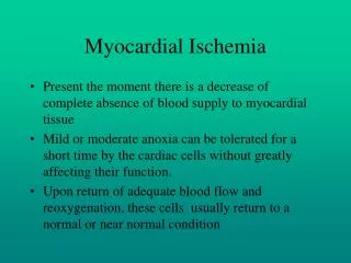
Epidemiology Cardiomyopathy is an important cause of morbidity and mortality among the world's aging population. Extension of DCMP varies over the range from 5 to 10 cases to 100 000 of population, it attacks men 25-45 years old more frequent.
Etiology Despite the fact that etiology of DCMP doesn’t determined in the scientific literature it is discussed about the connection of it with myocarditis (especially virus), myocardium damage by toxins (such as ethanol, cobalt, anthracyclins), endocrine diseases (pathology of thyroid gland, hypophisis) and some metabolic disturbances. Probably, genetic factors, especially X-chromosome-defect, take part.
Pathogenesis Degeneration and downfall of cardiomyocytes with stromal collapse occur under influences of unknown factor. It results in losing of ability to resist to expansion. Then interstitial and perivascular myocardial fibrosis, hypertrophy of undamaged muscular fibers, dilatation of heart cavities, cardiomegaly, severe arrhythmias, heart failure are formed.
Pathological anatomy Reliable pathomorphological hallmark of DCMP is dilatation of heart cavities under the absence of coronary atherosclerosis. In 50% of cases in dilated heart cavities thrombi can be found. Myocardium is pale and limp. Findings may include myocardial injury with inflammatory mediators. Physical disruption of myocytes by inflammatory cells, proliferation of interstitial cells, and increased fibrous matrix may also be found.
DCMP are classified on: • promptly progressive, • slowly progressive, • stable.
Clinical features of DCMP are • progressive heart failure, • arrhythmias, • thromboembolism, • sudden death, • cardiomegaly, • systolic murmur of the mitral regurgitation.
Additional methods of research Laboratory studies. There are no specific signs for DCMP
ECG • nonspecific ST segment and T wave changes. • different arrhythmias and blocks. • left ventricular hypertrophy or other chamber enlargement are observed on ECG. The most often ECG features are atrial fibrillation and left bundle-branch block.
X-ray Cardiomegaly which can be observed at X-ray examination is one of the most sensitive and specific signs in diagnosing of this cardiomyopathy.
Echocardiography Globally dilated hypokinetic heart with low ejection fraction can be determined. Mitral and tricuspid regurgitation, LV mural thrombus are common for dilated cardiomyopathy.
Other methods • coronarography, • scintigraphy, • ventriculography, • heart catheterization, • endomyocardial biopsy can be used in differential diagnostics
Diagnosis Diagnosis is lawful if patient has got heart failure with dilated heart and there are no other diseases which could cause such dilatation.
Differential diagnosis You must differentiate DCMP with • myocarditis, • IHD, • congenital and acquired heart diseases, • pericarditis, • heart swellings, • hypertrophic cardiomyopathy, • restrictive cardiomyopathy.
Management Dietary recommendations include sodium and water restrictions. You can treat with • ACE-inhibitors (enalapril, monopril), • digoxin, • β-blockers (carvedilol, nebibolol), • aldosteron agonists (inspra, verospiron), • diuretics, • anticoagulants and antiagregants. • It consider heart transplantation.
Prognosis Patients with severe heart failure have more than a 50% yearly mortality rate. Patients with mild heart failure have significantly better prognoses, especially with optimal medical therapy. Prophylaxis.As etiology of this disease is unknown there are no measures of prophylaxis.
Hypertrophic cardiomyopathy (HCM) Definition. Hypertrophic cardiomyopathy is characterized by left ventricular hypertrophy, which is more often asymmetric and mainly involves the interventricular septum.
Epidemiology HCM is reported in 0.5% of the outpatient population referred for echocardiography. Peak of morbidity is the age 25-35 years old.
Etiology The molecular basis for HCM defects located in several of the genes encoding for the myosin, actin and tropomyosin. It can be related to endocrine abnormalities and sympathicoadrenal dysfunction.
Pathogenesis Hypertrophy of left ventricle makes myocardium rigid and causes trouble to diastoliс fillingthat lead to diastolic dysfunction and coronary deficiency. The hypertrophy of the interventricular septum results in an obstruction of left ventricular outflow tract. The pressure gradient increases due to systolic anterior motion of the mitral valve towards the hypertrophied septum.
Pathological anatomy Myocardial hypertrophy, disorganization of myofibrillar architecture and fibrosis occur in almost all patients with HCM. Besides, abnormal intramural coronary arteries, with a reduction in the size of the lumen and thickening of the vessel wall, are common in these patients. There are fibrosis and sclerosis of anterior mitral valve flapper.
Classification HCM can be subdivided into • obstructive (with pressure gradient between the aorta and LV on the level of its outflow tract), • nonobstructive (without pressure gradient) types.
Clinical features Symptoms can include • syncope and presyncope, • angina pectoris and • congestive heart failure. Impaired diastolic relaxation and markedly increased myocardial oxygen consumption are caused by ventricular hypertrophy that results in subendocardial ischemia, particularly during exertion.
Syncope Syncope as a rule, appear after physical exertion when degree of obstruction increase (preloadand afterload decrease, high level of catecholamine, which have positive inotropic effect, can be observed), аnd after peripheric vasodilatators, diuretics intake, (preloadand afterload decrease) and digoxin (due to their positive inotropic effect). Alternatively, syncope may be caused by arrhythmias, either tachycardias or bradycardias.
Angina pectoris Angina pectoris is caused by big myocardial mass, relative coronary deficiency and high pressure in left ventricle that compressed subendocardial vessels.
Congestive heart failure It may be a result of an impaired diastolic function. Systolic function is almost always well preserved at these patients.
Other clinical features • Systolic ejection murmur is the best heard between the apex and left sternal border and radiates to the suprasternal notch but not to the carotid arteries or neck and increase in vertical position. • Apex beat is displacedleft due to heart hypertrophy.
Additional methods of researches Lab Studies: No specific laboratory blood tests are required in the workup of HCM. Genetic testing can help in making diagnosis of HCM.
Electrocardiography Common findings include • abnormalities of segment ST and T-wave, • LV hypertrophy.
Electrocardiography Other often findings observed on ECG include • conduction abnormalities, • and ventricular arrhythmias.
Chest X-ray Enlargement of left ventricle can be observed often on chest X-ray.
Echocardiography finds • LV, especially septal hypertrophy; • Systolic anterior motion of the anterior mitral valve leaflet; • The hallmark of obstructive HCM is a pressure gradient between the aorta and LV on the level of its outflow tract; • Diastolic dysfunction.
Diagnosis Presence of causeless myocardial hyperthrophy especially septum with signs of outflow-tract obstruction is quite enough for basing of diagnosis HCM.
Differential diagnosis HCM can be differentiated with diseases at which systolic murmur (aortic stenosis, mitral insufficiency, congenital heart abnormalities) and LV hypertrophy. Angina, arterial hypertension can be discussed.
Treatment First of all beta-blockers are used.They reduce inotropic function of left ventricle, decrease diastolic dysfunction, increase LV compliance and decrease pressure gradient across LV outflow tract, heart rate, myocardial oxygen consumption and cause reducing myocardial ischemia.
Treatment If Beta-blockers are contraindicated amiodaron or calcium antagonists verapamil group should be recommended. Sometimes antiarrhythmic drugs - Disopyramide and Sotalol - are used. Avoid inotropic drugs (glycosides, sympathomimetics), vasodilators and diuretics if possible. They could increase obstruction degree in outflow tract.
Surgical methods of treatment • Septal myomectomy, mitral valve replacement, selective arterial ethanol infusion (for damage of septum) could be used for treatment of HCM. • The implantable cardioverter defibrillator is used for the prevention of sudden arrhythmic death.
Prognosis Mortality rate is 4% per year. Sudden death is the most common reason. Prophylaxis There is no special prophylaxis at present because of absence knowledge of etiology of HCM. You should take care about prevention of IE at surgical operation. Patients must abstain from highly strenuous physical exertion.
Restrictive Cardiomyopathy (RCM) Definition. Restrictive Cardiomyopathy is a myocardial disease characterized by endocardial, subendocardial and myocardial fibrosis of ventricles.
Epidemiology It takes 5% of other cardiomyopathies but it is more often in the tropic countries. Etiology Most of researchers connect development of this disease with eosinophilia. It is supposed that the main protein of grainless eosinophils has evident cardiac toxicity. Besides the role of other factors are under discussion.
Pathogenesis Endocardial, subendocardial and myocardial fibrosis makes heart walls more stiff that causes diastolic dysfunction and heart failure.
Pathological anatomy Thickness of ventricular walls is normal. Endocardial tissue is hard and thick, papillary muscles and chords are fibrosed, atria are often dilated, ventricular cavities are reduced. Thrombi and fibrous tissues can be also founded in heart cavities and they can cause ventricular obliteration.
Classification There are two types of RCM : • - witheosinophilia (Loeffler endocarditis) • - without eosinophilia (endomyocardial fibrosis).
Clinical features Patients usually complain on • signs of heart failure, • arrhythmias and • thromboembolic complications. There is no cardiomegaly.
Additional methods of researches Lab Studies Eosinophilia is often in complete blood count of such patients. Electrocardiography The findings on ECG may be different. The most often is atrial fibrillation.
Chest X-ray It typically shows normal heart size.
Echocardiography It shows normal systolic function, nonhypertrophied left ventricle and marked dilatation of both atria. Doppler echocardiogram shows features of restriction to diastolic filling. The ventricular cavity size may be normal or reduced. There are mural thrombus.
Diagnosis At RCMyou should observe: • Heart failure; • Unchanged heart size; • Diastolic dysfunction; • The absence of diseases which can cause a such dysfunction.